. 2015 Apr;52(4):409-17.
doi: 10.1165/rcmb.2014-0312MA.

Isolation of epithelial, endothelial, and immune cells from lungs of transgenic mice with oncogene-induced lung adenocarcinomas

Affiliations

Isolation of epithelial, endothelial, and immune cells from lungs of transgenic mice with oncogene-induced lung adenocarcinomas

Amlak Bantikassegn et al. Am J Respir Cell Mol Biol. 2015 Apr.

Abstract

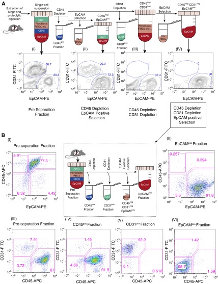
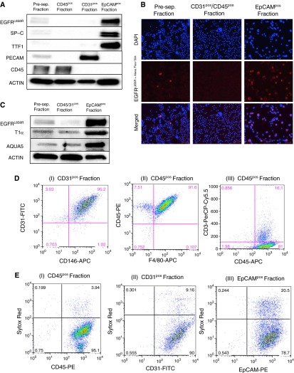
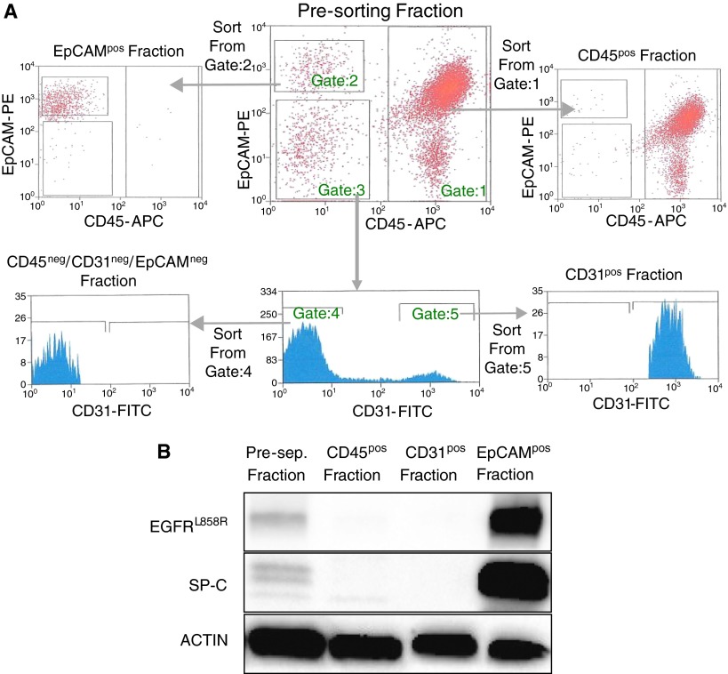
Genetically engineered mouse models of lung adenocarcinoma have proven invaluable for understanding mechanisms of tumorigenesis, therapy response, and drug resistance. However, mechanistic studies focused on studying these processes in tumor-bearing mouse lungs are confounded by the fact that, in most cases, relevant signaling pathways are analyzed in whole-lung preparations, which are composed of a heterogeneous mixture of cells. Given our increasing knowledge about the roles played by different subpopulations of cells in the development of lung adenocarcinoma, separating the major cellular compartments of the tumor microenvironment is recommended to allow for a precise analysis of relevant pathways in each isolated cell type. In this study, we optimized magnetic- and fluorescence-based isolation protocols to segregate lung epithelial (CD326/epithelial cell adhesion molecule-positive), endothelial (CD31-positive), and immune (CD45-positive) cells, with high purity, from the lungs of transgenic mice with mutant epidermal growth factor receptor-induced lung adenocarcinomas. This approach, which can potentially be extended to additional lung adenocarcinoma models, enables delineation of the molecular features of individual cell types that can be used to gain insight into their roles in lung adenocarcinoma initiation, progression, and response to therapy.

Keywords: epithelial cell adhesion molecule; epithelial cell isolation; lung adenocarcinoma; transgenic mouse models.


Figures

Figure 1.
Figure 1.
Optimization of the magnetic-activated cell sorting (MACS) protocol for isolating lung epithelial, endothelial, and immune cells. (A) Flow cytometry analysis of single-cell suspensions from the lungs of a tumor-bearing Clara cell secretory protein (CCSP)reverse tetracycline transactivator (rtTA) (+) tetracycline-responsive element (TetO) EGFRL858R (+) mouse (A [I]) before performing MACS-based separations. The whole single-cell suspension was then depleted of CD45pos cells, followed either directly by epithelial cell adhesion molecule (EpCAM)–positive selection (A [II]) or CD31 depletion (A [III and IV]). EpCAM-positive selection was then performed on a subset of the CD45negCD31neg cells (A [IV]). The highest epithelial cell purity was achieved using the latter protocol. All the fractions analyzed were gated on CD45neg cells. (B) Flow-based analysis of the EpCAMpos, CD31pos, and CD45pos fractions (B [II, IV–VI]) isolated using sequential CD45, CD31, and EpCAM selection (see center panel) compared with the preseparation fraction (B [I and III]). Flow analysis was performed on a FACSCalibur using CD31-fluorescein isothiocyanate (FITC), CD45-allophycocyanin (APC), and EpCAM-phycoerythrin (PE) antibodies. EGFR, epidermal growth factor receptor; Fib, fibroblasts; RBC, red blood cell.
Figure 2.
Figure 2.
Molecular and cellular characterization of the MACS-isolated cell factions. (A) Western blot analysis on MACS-isolated cells from an EGFRL858R tumor-bearing mouse for surfactant protein (SP)-C, EpCAM, thyroid transcription factor-1 (TTF1), CD45, platelet endothelial cell adhesion molecule (PECAM), and EGFRL858R, as indicated. (B) Immunocytochemistry for EGFRL858R in preseparation cells, in EpCAMpos cells, and in CD31pos/CD45pos cells. Cells were incubated with an EGFRL858R primary antibody (red), followed by a secondary antibody (Alexa Fluor 594) and 4′,6-diamidino-2-phenylindole (DAPI) (blue) before being imaged (scale bars, 50 μm). (C) Lysates from cells isolated from an EGFRL858R tumor-bearing mouse probed with antibodies to aquaporin 5 (AQUA5) and T1α (alveolar type I cell markers) are shown. Of note, the center lane shows lysates of cells after CD31/CD45 simultaneous depletion (not sequential). (D) Flow cytometry analysis of isolated nonepithelial cell fractions, as indicated, to confirm their identity. (E) Sytox red staining on MACS-isolated CD45-, CD31-, and EpCAM-expressing cells from tumor-bearing (EGFRL858R) mice, as indicated. The fractions were stained with Sytox red to visualize the proportion of viable cells. Of note, the EpCAMpos fraction was isolated using CD31/CD45 simultaneous depletion (not sequential) before the EpCAM-positive selection to maximize viability.
Figure 3.
Figure 3.
Fluorescence-activated cell sorting (FACS)–based isolation of EpCAMpos, CD45pos, and CD31pos cells from tumor-bearing (EGFRL858R) mouse lungs. (A) Cells from a single-cell suspension of lung tissue were labeled with EpCAM-PE, CD31-FITC, and CD45-APC before being sorted using the BD FACSAria. Cells were gated as follows: CD45-APC–expressing cells (Gate:1), EpCAM-PE expressing cells (Gate:2), and EpCAM-PE/CD45APC double-negative cells (Gate:3). To isolate the CD31-FITC–expressing cells, we visualized the EpCAM-PE/CD45-APC double-negative cells on a CD31-FITC histogram and identified cells positive for CD31-FITC (Gate:5) separately from the cells that were CD31-FITC, EpCAM-PE, and CD45-APC triple negative (Gate:4). Therefore, to isolate CD45pos, EpCAMpos, CD31pos, and triple-negative cells, we sorted from gates 1, 2, 5, and 4, respectively. (B) Western blot analysis on FACS-isolated cells from an EGFRL858R tumor-bearing mouse for SP-C and EGFRL858R.

References

    1. Reynolds SD, Malkinson AM. Clara cell: progenitor for the bronchiolar epithelium. Int J Biochem Cell Biol. 2010;42:1–4. - PMC - PubMed
    1. Broeckaert F, Bernard A. Clara cell secretory protein (CC16): characteristics and perspectives as lung peripheral biomarker. Clin Exp Allergy. 2000;30:469–475. - PubMed
    1. Ward HE, Nicholas TE. Alveolar type I and type II cells. Aust N Z J Med. 1984;14:731–734. - PubMed
    1. Fehrenbach H. Alveolar epithelial type II cell: defender of the alveolus revisited. Respir Res. 2001;2:33–46. - PMC - PubMed
    1. Phelps DS, Floros J. Localization of pulmonary surfactant proteins using immunohistochemistry and tissue in situ hybridization. Exp Lung Res. 1991;17:985–995. - PubMed

Publication types

MeSH terms