. 2015 Aug 6;34(32):4278-84.
doi: 10.1038/onc.2014.354. Epub 2014 Oct 27.

The impact of osteoblastic differentiation on osteosarcomagenesis in the mouse

Affiliations

The impact of osteoblastic differentiation on osteosarcomagenesis in the mouse

T Quist et al. Oncogene. .

Abstract

Osteosarcomas remain an enigmatic group of malignancies that share in common the presence of transformed cells producing osteoid matrix, even if these cells comprise a minority of the tumor volume. The differentiation state of osteosarcomas has therefore become a topic of interest and challenge to those who study this disease. In order to test how the cell of origin contributes to the final state of differentiation in the transformed cells, we compared the relative tumorigenicity of Cre-LoxP conditional disruption of the cell cycle checkpoint tumor-suppressor genes Trp53 and Rb1 using Prx1-Cre, Collagen-1α1-Cre and Osteocalcin-Cre to transform undifferentiated mesenchyme, preosteoblasts and mature osteoblasts, respectively. The Prx1 and Col1α1 lineages developed tumors with nearly complete penetrance, as anticipated. Osteosarcomas also developed in 44% of Oc-Cre;Rb1(fl/fl);Trp53(fl/fl) mice. We confirmed using 5-ethynyl-2'-deoxyuridine click chemistry that the Oc-Cre lineage includes very few actively cycling cells. By assessing radiographic mineralization and histological osteoid production, the differentiation state of tumors did not correlate with the differentiation state of the lineage of origin. Some of the osteocalcin-lineage-derived osteosarcomas were among the least osteoblastic. Osteocalcin immunohistochemistry in tumors correlated well with the expression of DNA methyl transferases, suggesting that silencing of these epigenetic regulators may influence the final differentiation state of an osteosarcoma. Transformation of differentiated, minimally proliferative osteoblasts is possible but may require such an epigenetic reprogramming that the tumors no longer resemble their differentiated origins.


Figures
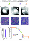
Figure 1
Figure 1. Osteosarcomagenesis can be induced by conditional tumor suppressor disruption across the range of osteoblast differentiation
(A) Schematic representing the differentiation course from mesenchymal stem cell to mature osteoblast with the expression point for each Cre-recombinase promoter plotted. (B) Representative radiographs and hematoxylin and eosin (H&E) histology photomicrographs from the osteosarcoma-mimicking skeletal tumors that arise following homozygous conditional disruption of floxed alleles of Trp53 and Rb1 induced by Prx1-Cre, Col1α1-Cre, or Oc-Cre. (C) Fraction of mice in each lineage that developed skeletal osteosarcoma-like tumors from a denominator sample size of 23, 32, and 25 mice for Prx1-Cre, Col1α1-Cre, and Oc-Cre, respectively. (D) Kaplan-Meier plots of the same cohorts presenting the relative latency to osteosarcomagenesis; censored animals reached morbidity requiring euthanasia without the development of a skeleton-associated tumor.
Figure 2
Figure 2. Active cell cycling among distinct osteoblast differentiation-state lineages
(A) Fluorescence photomicrographs of limb tissues from mice bearing a conditional tdTomato linage marker (red) activated by Prx1-Cre, Col1α1-Cre, or Oc-Cre, collected two days after the reported expression of each and 4 hours after intraperitoneal administration of 50 mg/kg EdU. Cells in active S-phase during that four hour period were detected by EdU Click-iT chemistry detection kit (Life Technologies) on 8μm frozen sections obtained after embedding in OCT (Tissue-Tek, Sakura Finetek). (B) Fraction of cells in each tdTomato-fluorescent lineage that co-stained with EdU as evidence of active cell cycling based off of 3 Prx1-Cre, 6 Col1α1-Cre, and 16 Oc-Cre tissue samples analyzed with ImageJ software.
Figure 3
Figure 3. Radiographic mineralization and osteoid matrix production do not correlate with the differentiation state of an osteosarcoma’s cell of origin
(A) Representative radiographs for each class of differentiation (1 through 4) as assessed by tumor mineralization. Lower and higher classes associated with lower and higher mineralized osseous matrix relative to the femoral diaphysis, respectively, in the tumors. (B) Distribution of radiographic mineralization classes among 48 Prx1-Cre;Rb1fl/fl;Trp53fl/fl, 48 Col1α1-Cre;Rb1fl/fl;Trp53fl/fl, and 18 Oc-Cre;Rb1fl/fl;Trp53fl/fl tumors. (C) Representative hematoxylin and eosin (H&E) photomicrographs of sectioned tumors demonstrating areas with the range of classes (1 through 4) of osteoid matrix production within tumors. Lower and higher classed areas associated with lower and higher production of osteoid matrix, respectively, in the tumors. (D) Distribution (and mean for each group, denoted by the +) of osteoid matrix production classes among skeletal tumors arising in 8 Prx1-Cre;Rb1fl/fl;Trp53fl/fl, 9 Col1α1-Cre;Rb1fl/fl;Trp53fl/fl, and 6 Oc-Cre;Rb1fl/fl;Trp53fl/fl mice. Tumor class, based on mid-cross-sections of tumors, was determined via an average of each class weighted by its associated cross-sectional area measured by ImageJ.
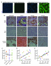
Figure 4
Figure 4. Dedifferentiation occurs during osteosarcomagenesis and correlates with silencing of epigenetic regulators
(A) Immunofluorescence photomicrographs of representative low matrix- and high matrix-producing osteosarcomas from Oc-Cre;Rb1fl/fl;Trp53fl/fl mice demonstrating loss (left) and retention (right) of Cre-recombinase, following 10mM sodium citrate boiling antigen retrieval for 15 minutes, staining with an anti-Cre primary antibody (Covance, 1:3000) followed by an Alexa 488 secondary antibody (Jackson, 1:500). (B) Representative photomicrographs from immunohistochemical stains with anti-osteocalcin (Abcam, 1:500 dilution, following 20 minute protease 2 treatment, but no other antigen retrieval), anti-Dnmt1 (LifeSpan Biosciences, 1:200 dilution), anti-Dnmt3a (Novus Biologicals, 1:50 dilution), and anti-Dnmt3b (Novus Biologicals, 1:100 dilution) antibodies, applied each for 2 hours at 37°C, following 1-hour pH8.0 cell conditioning 1 (Ventana Medical Systems) antigen retrieval, using the BenchMark Ultra automated immunostainer (Ventana Medical Systems) with a biotinylated secondary antibody, DAB detection (IView) and hematoxylin counterstain. (C) Osteocalcin immunohistochemical ranking by 2 investigators blinded to sample identification, charted against Cre-initiation group, demonstrating no correlation. (D) Chart of osteocalcin rank by osteoid matrix production rank, demonstrating modest correlation. (E) Mean Dnmt immunohistochemical rank, averaged from individual Dnmt1, Dntm3a, and Dnmt3b ranks, charted by osteocalcin rank. (All magnification bars = 10μm)

References

    1. Dick JE. Breast cancer stem cells revealed. Proceedings of the National Academy of Sciences of the United States of America. 2003;100(7):3547–9. - PMC - PubMed
    1. Reya T, Morrison SJ, Clarke MF, Weissman IL. Stem cells, cancer, and cancer stem cells. Nature. 2001;414(6859):105–11. - PubMed
    1. Sharkey FE. Morphometric analysis of differentiation in human breast carcinoma. Tumor heterogeneity. Archives of pathology & laboratory medicine. 1983;107(8):411–4. - PubMed
    1. Meacham CE, Morrison SJ. Tumour heterogeneity and cancer cell plasticity. Nature. 2013;501(7467):328–37. - PMC - PubMed
    1. Angus-Hill ML, Elbert KM, Hidalgo J, Capecchi MR. T-cell factor 4 functions as a tumor suppressor whose disruption modulates colon cell proliferation and tumorigenesis. Proceedings of the National Academy of Sciences of the United States of America. 2011;108(12):4914–9. - PMC - PubMed

Publication types

MeSH terms