. 2015 Jan 15;21(2):396-404.
doi: 10.1158/1078-0432.CCR-14-1591. Epub 2014 Oct 27.

Combined MEK and PI3K inhibition in a mouse model of pancreatic cancer

Affiliations

Combined MEK and PI3K inhibition in a mouse model of pancreatic cancer

Brinda Alagesan et al. Clin Cancer Res. .

Abstract

Purpose: Improved therapeutic approaches are needed for the treatment of pancreatic ductal adenocarcinoma (PDAC). As dual MEK and PI3K inhibition is presently being used in clinical trials for patients with PDAC, we sought to test the efficacy of combined targeting of these pathways in PDAC using both in vitro drug screens and genetically engineered mouse models (GEMM).

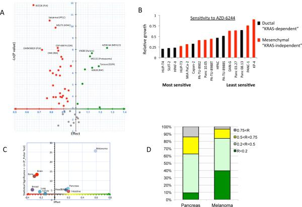
Experimental design: We performed high-throughput screening of >500 human cancer cell lines (including 46 PDAC lines), for sensitivity to 50 clinically relevant compounds, including MEK and PI3K inhibitors. We tested the top hit in the screen, the MEK1/2 inhibitor, AZD6244, for efficacy alone or in combination with the PI3K inhibitors, BKM120 or GDC-0941, in a Kras(G12D)-driven GEMM that recapitulates the histopathogenesis of human PDAC.

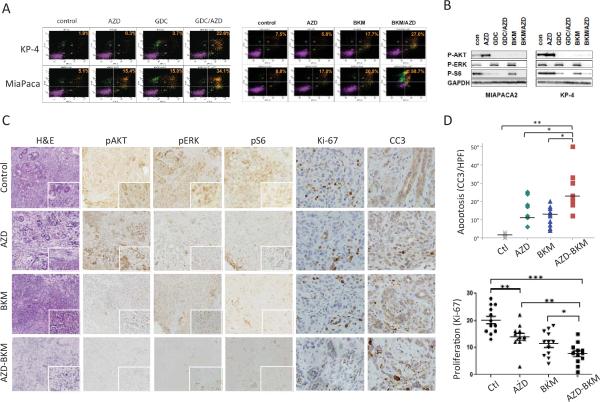
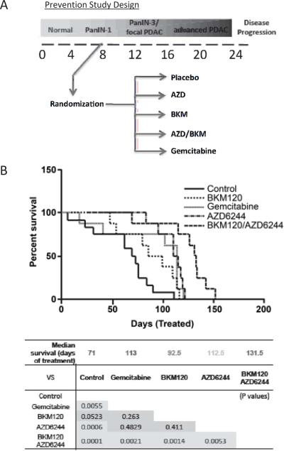
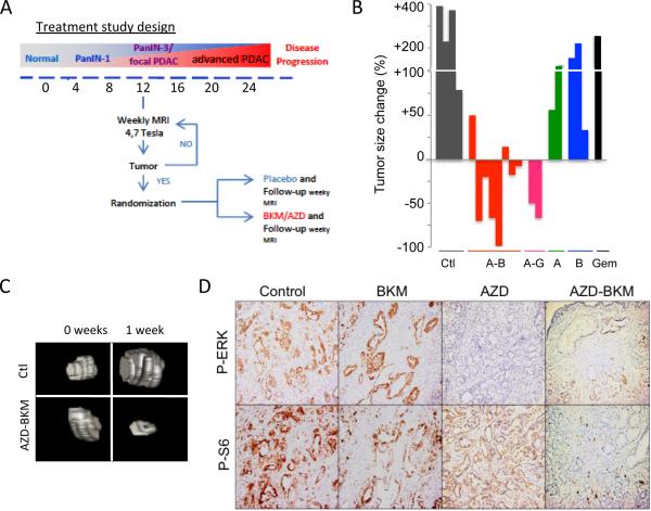
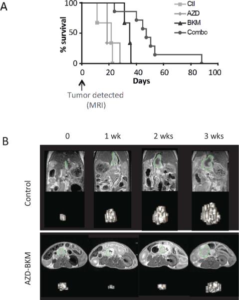
Results: In vitro screens revealed that PDAC cell lines are relatively resistant to single-agent therapies. The response profile to the MEK1/2 inhibitor, AZD6244, was an outlier, showing the highest selective efficacy in PDAC. Although MEK inhibition alone was mainly cytostatic, apoptosis was induced when combined with PI3K inhibitors (BKM120 or GDC-0941). When tested in a PDAC GEMM and compared with the single agents or vehicle controls, the combination delayed tumor formation in the setting of prevention and extended survival when used to treat advanced tumors, although no durable responses were observed.

Conclusions: Our studies point to important contributions of MEK and PI3K signaling to PDAC pathogenesis and suggest that dual targeting of these pathways may provide benefit in some patients with PDAC. Clin Cancer Res; 21(2); 396-404. ©2014 AACR.


Figures

Figure 1
Figure 1. High-throughput screening identifies the MEK1/2 inhibitor, AZD6244 as the most active compound against PDAC cell lines
A) Volcano plot representing the responsiveness of PDAC cell lines (N=46) compared to non-PDAC cancer cell lines (N=500) to a series of 50 potential anti-cancer drugs. X axis: relative sensitivity (Effect > 1: PDAC cells are on average more sensitive than non-PDAC cells), Y axis: statistical significance (Gray dots: compounds with p>0.05; Green dots: compounds preferentially targeting PDAC; Red dots: compounds for which PDAC cell lines are more resistant than other types of cell lines). B) Relative sensitivity to AZD-6244 of PDAC cell lines identified as “KRAS-dependent” and “–independent”(8, 9). C) Organ specificity profile of AZD-6244. Cell lines from cancer types were iteratively compared to the rest of the cell collection to determine the specific targeting by tissue of origin. X axis: relative sensitivity (Effect: average survival of cell lines from a given organ/survival of the other lines). Y axis: statistical significance of the organ enrichment (Fisher exact test). D) Relative sensitivity of PDAC and melanoma lines to 2 μM AZD-6244. Bar color indicates relative cell number (R) compared to control. Bar height represents the percentage of cell lines of each tumor type showing the indicated degree of growth inhibition.
Figure 2
Figure 2. Analysis of cell lines and ex vivo organotypic cultures supports the combined targeting of MEK and PI3K in PDAC
A, B) Treatment of PDAC cell lines with vehicle control or with the indicated drugs (AZD: AZD-6244; BKM: BKM-120; GDC: GDC-0941) used at 1 μ#. A) FACS plots showing PI/AnnexinV staining. The % of apoptotic cells is indicated. B) Western blot showing effect of the inhibitors on p-AKT (Thr308) and p-ERK (Thr202/Tyr204) levels. C-D) Analysis of therapeutic responses of ex vivo organotypic cultures of primary PDAC. C) Freshly derived organotypic cultures were treated with the indicated compounds (each at 1 μM) for 24 hours and then processed for staining with H&E or with antibodies to p-ERK (Thr202/Tyr204), p-AKT (Thr308), p-S6 (Ser235/236), Ki-67, and cleaved Caspase 3. D) Quantification of apoptosis at 24 hrs (cleaved caspase-3 staining) and of proliferation (Ki-67 staining). Statistical significance is indicated; p<0.01 (*), p<0.0001 (**).
Figure 3
Figure 3. Combined targeting of MEK and PI3K delays the progression of PDAC from PanIN lesions in the KRAS-p53 mouse model
A) Schematic of experimental design for prevention studies. Mice were treated prior to the onset of PDAC and monitored for evidence of tumor progression. B) Survival curve (Kaplan-Meier Analysis) calculated as length of time between start of treatment and sacrifice. Chart shows statistical analysis of survival data.
Figure 4
Figure 4. Advanced PDAC in the KRAS-p53 mouse model are responsive to dual MEK/PI3K inhibition
A) Schematic of experimental design for treatment of advanced PDAC. Mice were monitored for the presence of tumors by MRI. Upon detection of PDAC 3-10 mm in size, mice were randomized into the treatment and control groups. B) Waterfall plot showing efficacy of AZD-6244 (A) combined with either BKM-120 (B) or GDC-0941 (G) in promoting PDAC regression. No responses were seen with the single agents or with gemcitabine (Gem). Statistical significance: combination versus control (p<0.0001), versus AZD-6244 (p<0.01), versus BKM-120 (p<0.001). C) Representative three-dimensional reconstructions of MRI scans prior to treatment, or after 1 week in the indicated treatment groups. D) Immunohistochemical staining for p-ERK (Thr202/Tyr204) and p-S6 (Ser235/236), in PDAC isolated from KRAS-p53 mice treated for 1 week with the indicated compounds.
Figure 5
Figure 5. Clinical benefit of dual MEK/PI3K inhibition in the KRAS-p53 PDAC models
A) Survival curve (Kaplan-Meier analysis) of KRAS-53 mice after MRI detection of PDAC and administration of the indicated treatments. The differences between the AZD-6244/BKM-120 combination and the other groups are statically significant (compared to control, p=0.0008; compared to AZD, p=0.0061; compared to BKM, p=0.022). B) Representative MRI scans of KRAS-p53 mice and 3D reconstructions at sequential time points in the indicated treatment groups. The vehicle-treated mouse show rapid tumor progression (upper panel). Treatment with AZD-6244 and BKM-120 results in partial tumor regression in some mice, although the effects are transient.

Similar articles

Cited by

References

    1. Siegel R, Ma J, Zou Z, Jemal A. Cancer statistics, 2014. CA: a cancer journal for clinicians. 2014;64:9–29. - PubMed
    1. Hidalgo M. Pancreatic cancer. N Engl J Med. 2010;362:1605–17. - PubMed
    1. Conroy T, Desseigne F, Ychou M, Bouche O, Guimbaud R, Becouarn Y, et al. FOLFIRINOX versus gemcitabine for metastatic pancreatic cancer. N Engl J Med. 2011;364:1817–25. - PubMed
    1. Moore MJ, Goldstein D, Hamm J, Figer A, Hecht JR, Gallinger S, et al. Erlotinib plus gemcitabine compared with gemcitabine alone in patients with advanced pancreatic cancer: a phase III trial of the National Cancer Institute of Canada Clinical Trials Group. J Clin Oncol. 2007;25:1960–6. - PubMed
    1. Feig C, Gopinathan A, Neesse A, Chan DS, Cook N, Tuveson DA. The pancreas cancer microenvironment. Clinical cancer research : an official journal of the American Association for Cancer Research. 2012;18:4266–76. - PMC - PubMed

Publication types