. 2015 Jan;35(1):199-210.
doi: 10.1128/MCB.00981-14. Epub 2014 Oct 27.

A novel role for protein kinase Kin2 in regulating HAC1 mRNA translocation, splicing, and translation

Affiliations

A novel role for protein kinase Kin2 in regulating HAC1 mRNA translocation, splicing, and translation

Ashish Anshu et al. Mol Cell Biol. 2015 Jan.

Abstract

A signaling network called the unfolded protein response (UPR) resolves the protein-folding defects in the endoplasmic reticulum (ER) from yeasts to humans. In the yeast Saccharomyces cerevisiae, the UPR activation involves (i) aggregation of the ER-resident kinase/RNase Ire1 to form an Ire1 focus, (ii) targeting HAC1 pre-mRNA toward the Ire1 focus that cleaves out an inhibitory intron from the mRNA, and (iii) translation of Hac1 protein from the spliced mRNA. Targeting HAC1 mRNA to the Ire1 focus requires a cis-acting bipartite element (3'BE) located at the 3' untranslated leader. Here, we report that the 3'BE plays an additional role in promoting translation from the spliced mRNA. We also report that a high dose of either of two paralogue kinases, Kin1 and Kin2, overcomes the defective UPR caused by a mutation in the 3'BE. These results define a novel role for Kin kinases in the UPR beyond their role in cell polarity and exocytosis. Consistently, targeting, splicing, and translation of HAC1 mRNA are substantially reduced in the kin1Δ kin2Δ strain. Furthermore, we show that Kin2 kinase domain itself is sufficient to activate the UPR, suggesting that Kin2 initiates a signaling cascade to ensure an optimum UPR.


Figures

FIG 1
FIG 1
Nucleotides GG1143-1144 play an important role in regulating HAC1 mRNA targeting and splicing. (A) Schematic representation of the HAC1 pre-mRNA. The 7-methylguanosine (m7G) cap, 5′- and 3′UTRs, exons, intron, and polyadenine (An) tail are shown. The 3′-bipartite element (3′BE) is shown in a box. Two conserved RNA motifs (5′-U1141GGCGC1148-3′ in red and 5′-G1180CGAC1184-3′ in green) are predicted to interact with each other (12), thus forming a helix-bulge (HB) structure. The adenine of the start codon AUG is assigned position 1. Other nucleotides are numbered accordingly and shown on top of the sketch. (B) Mutations of the nucleotides GG1143-1144 impair yeast cell growth on the tunicamycin medium. The hac1Δ yeast strains expressing the indicated HAC1 alleles were serially diluted and spotted on SD medium alone and SD medium containing tunicamycin. (C) Mutations of the GG1143-1144 nucleotides reduce HAC1 mRNA splicing. Total RNA was isolated from yeast strains indicated in panel B, and RT-PCR was used to analyze spliced (HAC1s) and unspliced (HAC1u) HAC1 transcripts. The relative levels of HAC1s transcript were estimated by measuring the ratio of HAC1s and total (HAC1u plus HAC1s) band intensities (measured by NIH ImageJ software), normalizing the ratio with the respective intensity of the 28S rRNA band, and then turning the resulting ratio value into percentage. (D) The deletion of the 3′BE reduces the ER stress response. A hac1Δ ire1Δ pIRE1-YFP yeast strain expressing the indicated HAC1-NRE variant was tested for growth on SD and tunicamycin media. (E) Analysis of HAC1 mRNA transcripts. RT-PCR was used to analyze spliced (HAC1s) and unspliced (HAC1u) HAC1 transcripts in cells indicated in panel D. (F) The GG1143-1144CC mutations impair HAC1 mRNA colocalization with Ire1 protein. An ire1Δ hac1Δ strain coexpressing three plasmid-borne genes IRE1-YFP, the ND-GFP2 gene, and the indicated WT HAC1 or its derivative, HAC1-NRE-ΔBE or HAC1-NRE-GG1143-1144CC was grown under an ER stress condition. Ire1 (red dots) and HAC1 mRNA (green dots) foci were visualized by confocal microscopy.
FIG 2
FIG 2
The GG1143-1144 nucleotides promote translation of HAC1 mRNA. (A and B) The intronless HAC1 (HAC1i) variant exhibits a slow-growth phenotype. (A) An ire1Δ yeast strain containing an empty vector or the same vector bearing a wild-type HAC1 or HAC1i variant was tested for growth on SD and tunicamycin media. (B) A hac1Δ yeast strain containing an empty vector or the same vector expressing the indicated HAC1i gene or its derivatives was tested for growth on the SD medium. In the lower panels, total RNA was isolated from strains indicated in panel B, and RT-PCR was used to monitor the levels of HAC1 mRNA. (C) The GG1143-1144CC mutations in the HAC1i allele decrease the Hac1 protein production. The whole-cell extracts were subjected to Western blot (WB) analysis using an antibody raised against the recombinant Hac1 protein. (D) The GG1143-1144CC mutations reduce the Hac1-induced gene expression. The yeast strains indicated in panel B were transformed with a plasmid containing a lacZ reporter gene under the control of a UPR element from the yeast KAR2 gene (11). The β-galactosidase activities were monitored in those transformants in the presence (+) and absence (−) of DTT as described in Materials and Methods. The experiments were repeated three times, and the plotted histogram represents the mean fold change with standard errors.
FIG 3
FIG 3
Both Kin1 and Kin2 promote the Ire1/HAC1-mediated UPR. (A) Overexpression of Kin2 restores the UPR of the HAC1-GG1143-1144CC allele. (Left) A hac1Δ kin1Δ yeast strain was transformed with a 2μ URA3 vector or the same vector carrying a WT KIN2 gene. In the resultant transformant, the indicated WT HAC1, HAC1-G661C, or HAC1-GG1143-1144CC was introduced. Yeast cells were then tested for growth on SD and tunicamycin media (left panels), and RT-PCR was used to analyze HAC1u and HAC1s mRNA populations under a condition of ER stress (right panel). (B) Overexpression of Kin1 restores the UPR of the HAC1-GG1143-1144CC allele. (Left) An hac1Δ kin2Δ yeast strain was transformed with a 2μ URA3 vector or the same vector carrying a WT KIN1 gene. In the resultant transformant, the indicated WT HAC1, HAC1-G661C, or HAC1-GG1143-1144CC gene was introduced. Yeast cells were then tested for growth on SD and tunicamycin media (left panels), and RT-PCR was used to analyze the HAC1u and HAC1s mRNA populations under a condition of ER stress (right panel). (C) The Kin2 kinase domain function is essential for rescuing the UPR in HAC1-GG1143-1144CC cells. A hac1Δ kin1Δ pHAC1-GG1143-1144CC yeast strain was transformed with an empty vector and the same vector carrying the indicated WT Kin2 or Kin2-D248A mutant. The resultant strains were tested for growth on SD and tunicamycin media (left panels), and RT-PCR was used to analyze the level of the HAC1s mRNA population (right panel).
FIG 4
FIG 4
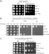
The kinase activity of Kin1 and Kin2 is required for the ER stress response. (A) The UPR is impaired in the kin1Δ kin2Δ strain. The WT yeast strain and its isogenic kin1Δ, kin2Δ, and kin1Δ kin2Δ mutants were grown, serially diluted, spotted, and tested for growth on SD and tunicamycin media. (B) Ire1, Hac1, and Kin1 Kin2 null mutant strains display differential sensitivities to tunicamycin. A yeast strain lacking ire1, hac1, or both kin1 and kin2 was grown, serially diluted (100 to 10−3), and spotted and tested for growth on both SD and tunicamycin media. (C) The kinase-dead mutants of Kin1 and Kin2 reduce the ER stress response. A kin1 kin2Δ yeast strain expressing a WT protein (Kin1 or Kin2) or its kinase-dead mutant (Kin1-D289A or Kin2-D248A) was tested for growth on both SD and tunicamycin media.
FIG 5
FIG 5
Both Kin1 and Kin2 modulate the Hac1-mediated ER stress response. (A and B) Both splicing and translation of HAC1 mRNA are reduced in the kin1Δ kin2Δ strain. (A) A wild-type (WT) strain or an isogenic kin1Δ kin2Δ mutant strain was grown in either the presence (+) or absence (−) of 20 mM DTT. Total RNA was isolated, and RT-PCR was used to analyze HAC1s and HAC1u mRNA transcripts. (B) Whole-cell extracts were subjected to Western blot (WB) analysis using an antibody against the Hac1 protein. (C) Expression of the Hac1-induced lacZ reporter is decreased in the kin1Δ kin2Δ strain. Expressions of lacZ were monitored in the presence (+) and absence (−) of DTT in the WT and kin1Δ in2Δ yeast strains, each bearing a UPRE-driven lacZ reporter gene.
FIG 6
FIG 6
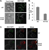
Kinases Kin1 and Kin2 play a role in focus formation as well as in colocalization of HAC1 mRNA with Ire1 protein. (A and B) The number of HAC1 mRNA foci is reduced in the kin1Δ kin2Δ strain. The indicated WT and kin1Δ kin2Δ yeast strains coexpressing the HAC1-NRE mRNA and the ND-GFP2 fusion protein were grown under an ER stress condition. (B) The HAC1 mRNA foci were counted using a confocal microscope. We chose those cells where at least one focus was observed. Approximately 60 cells were randomly selected and analyzed from 20 different microscopic fields of view. (C) Analysis of colocalization of HAC1 mRNA and Ire1 protein. The yeast cells indicated in panel A were transformed with an Ire1-YFP fusion protein (21). The HAC1 mRNA (green dots) and Ire1 protein (red dots) were visualized by confocal microscopy. Images were merged to show their colocalization (indicated by arrows).
FIG 7
FIG 7
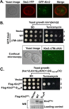
The kinase domain of Kin2 plays a role in the UPR. (A) Kin2 kinase is located in the cytoplasm: An ste2Δ yeast strain coexpressing Ste2-YFP and GFP-Kin2 fusion proteins was scanned using a confocal microscope. The Ste2-YFP (red circle) and GFP-Kin2 (green dots) fusion proteins were detected. (B) The deletion of both the predicted transmembrane (TM) domain and the kinase-associated domain 1 (KA1) does not impair Kin2 function. A kin1Δ kin2Δ yeast strain expressing GFP-Kin2 or its derivative GFP-Kin2-ΔKA1-ΔTM variant grew on the tunicamycin medium. (Lower panel) Confocal microscopy revealed that the GFP-Kin2-ΔKAI-ΔTM protein (green dots) displayed a punctate distribution. (C) The Kin2 kinase domain (Flag-Kin2KD) is sufficient to overcome the defective UPR of the HAC1-GG1143-1144CC cells. A hac1Δ kin1Δ pHAC1-GG1143-44CC yeast strain expressing Flag-Kin2KD or its derivative, Flag-Kin2KD-D248A, was tested for growth on the SD and tunicamycin media. (Lower panel) Whole-cell extracts from the indicated cells were subjected to Western blotting (WB) using an anti-Flag antibody. Nonspecific bands are shown as loading.
FIG 8
FIG 8
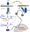
Model of activation of ER stress response by both Ire1 and Kin kinases. The ER-resident kinase/RNase Ire1 resides across the ER membrane. (Both the NH3+-terminal luminal and cytoplasmic kinase/RNase domains are colored teal.) Under conditions of ER stress, Ire1 cleaves out an intron (orange line) in the HAC1 pre-mRNA. The resulting mature mRNA translates the Hac1 protein (Hac1p) that activates transcription of the UPR genes in the nucleus. In parallel, the protein kinase Kin1 or Kin2 (shown by blue with both the NH3+-terminal and KA1 regions likely to anchor the endomembrane) is coactivated. There are two possible ways Kin1 and Kin2 functionally contribute to the HAC1-mediated UPR. First, Kin1/Kin2 binds directly to the 3′UTR element of HAC1 mRNA and promotes targeting to the ER stress signaling site, splicing, and translational repression. Second, Kin1/Kin2 phosphorylates a protein substrate (substrate shown by “Skin,” and phosphorylation is indicated by “P”), a component of the 3′UTR-RNP complex that modulates both mRNA targeting and translation. Alternatively, the Kin1/Kin2 substrate constitutes a novel signaling cascade (denoted by “?” in a box) and transactivates the UPR genes in the nucleus. The sensing mechanism for Kin2 is unknown and is shown by “?”

Similar articles

Cited by

References

    1. Hebert DN, Molinari M. 2007. In and out of the ER: protein folding, quality control, degradation, and related human diseases. Physiol Rev 87:1377–1408. doi:10.1152/physrev.00050.2006. - DOI - PubMed
    1. Ron D, Walter P. 2007. Signal integration in the endoplasmic reticulum unfolded protein response. Nat Rev Mol Cell Biol 8:519–529. doi:10.1038/nrm2199. - DOI - PubMed
    1. Walter P, Ron D. 2011. The unfolded protein response: from stress pathway to homeostatic regulation. Science 334:1081–1086. doi:10.1126/science.1209038. - DOI - PubMed
    1. Ellgaard L, Helenius A. 2003. Quality control in the endoplasmic reticulum. Nat Rev Mol Cell Biol 4:181–191. doi:10.1038/nrm1052. - DOI - PubMed
    1. Bernales S, Schuck S, Walter P. 2007. ER-phagy: selective autophagy of the endoplasmic reticulum. Autophagy 3:285–287. doi:10.4161/auto.3930. - DOI - PubMed

Publication types

MeSH terms

LinkOut - more resources