. 2014 Dec 12;289(50):34728-42.
doi: 10.1074/jbc.M114.585505. Epub 2014 Oct 27.

SCCRO3 (DCUN1D3) antagonizes the neddylation and oncogenic activity of SCCRO (DCUN1D1)

Affiliations

SCCRO3 (DCUN1D3) antagonizes the neddylation and oncogenic activity of SCCRO (DCUN1D1)

Guochang Huang et al. J Biol Chem. .

Abstract

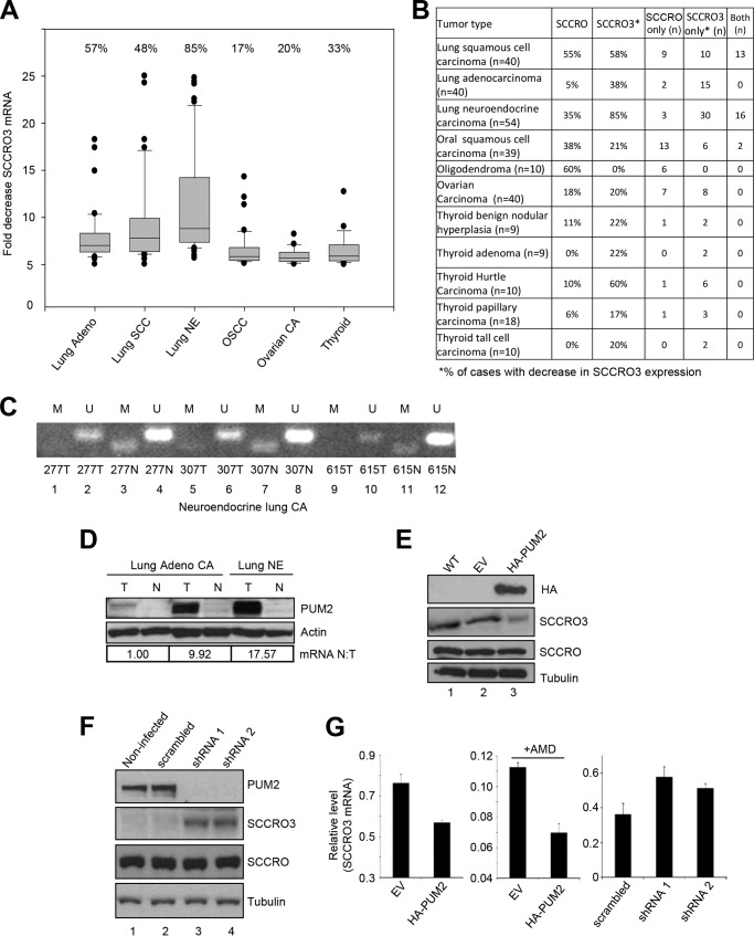
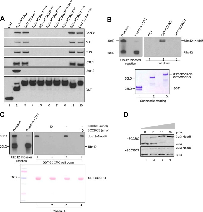
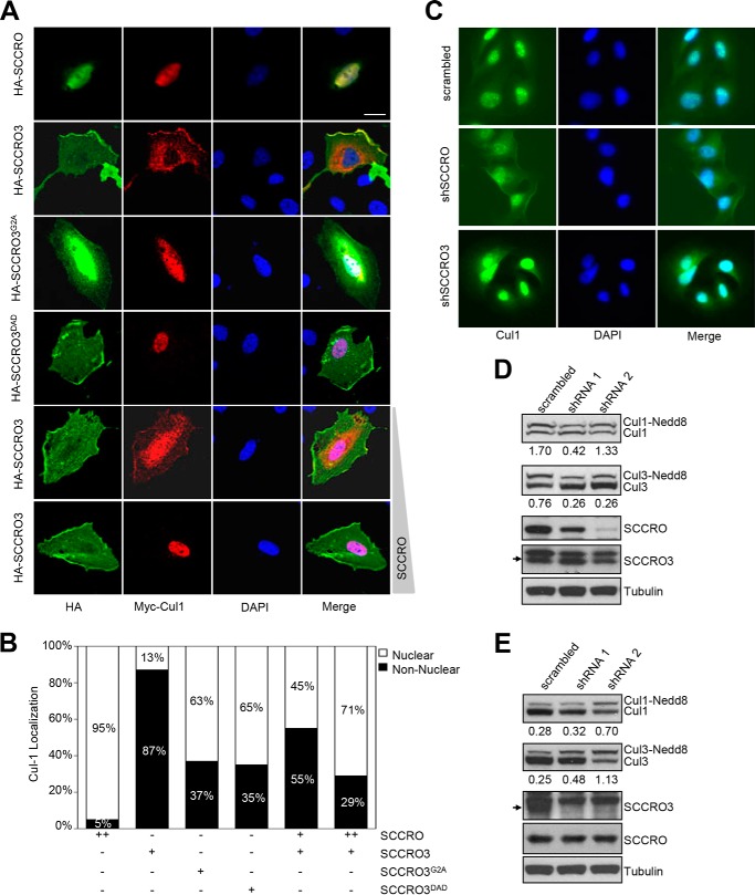
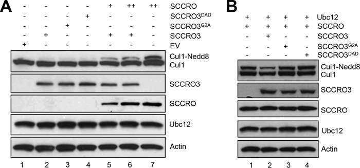
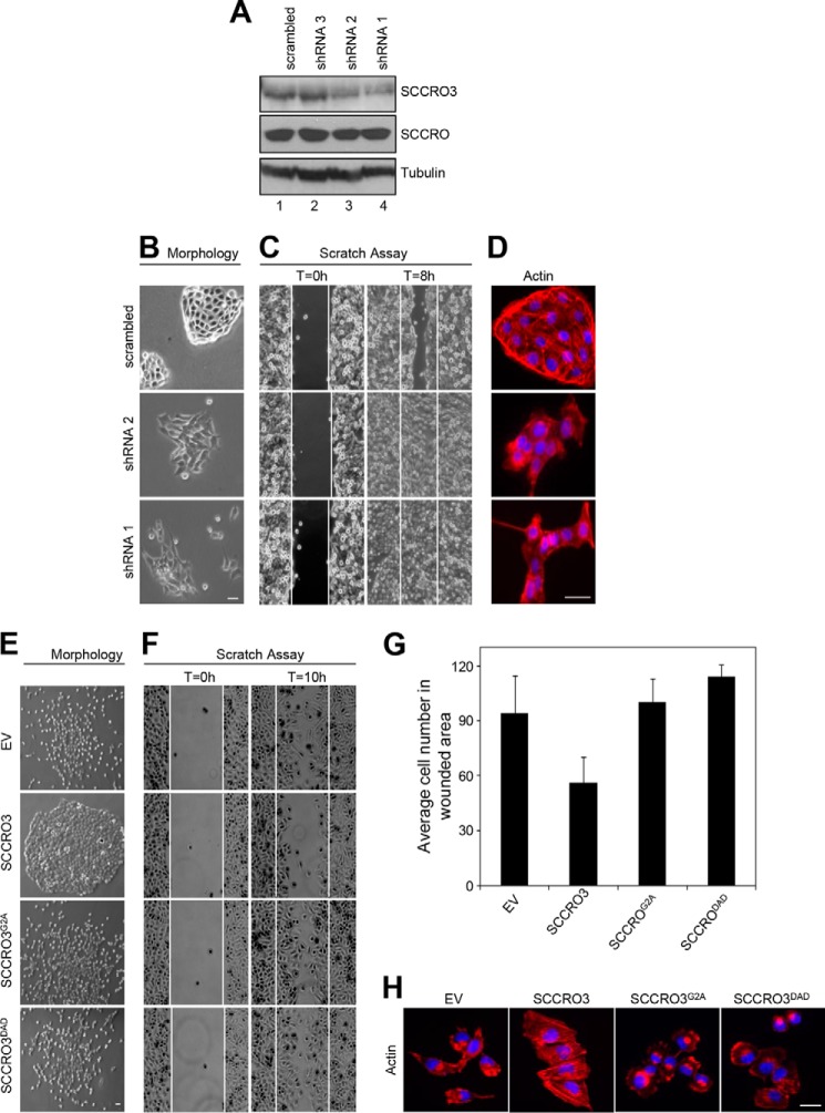
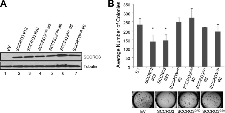
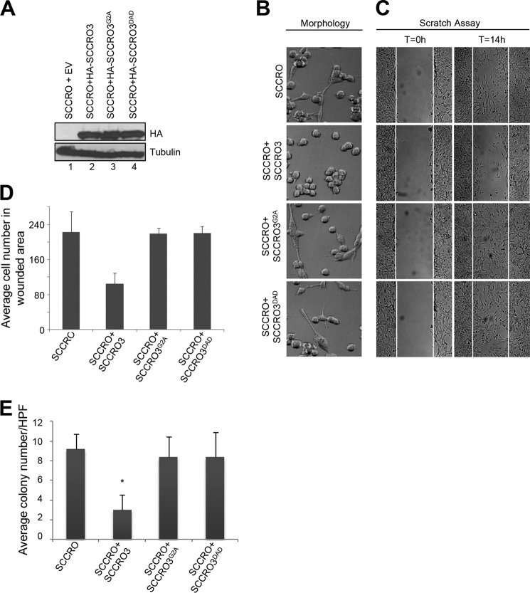
The activity of cullin-RING type ubiquitination E3 ligases is regulated by neddylation, a process analogous to ubiquitination that culminates in covalent attachment of the ubiquitin-like protein Nedd8 to cullins. As a component of the E3 for neddylation, SCCRO/DCUN1D1 plays a key regulatory role in neddylation and, consequently, cullin-RING ligase activity. The essential contribution of SCCRO to neddylation is to promote nuclear translocation of the cullin-ROC1 complex. The presence of a myristoyl sequence in SCCRO3, one of four SCCRO paralogues present in humans that localizes to the membrane, raises questions about its function in neddylation. We found that although SCCRO3 binds to CAND1, cullins, and ROC1, it does not efficiently bind to Ubc12, promote cullin neddylation, or conform to the reaction processivity paradigms, suggesting that SCCRO3 does not have E3 activity. Expression of SCCRO3 inhibits SCCRO-promoted neddylation by sequestering cullins to the membrane, thereby blocking its nuclear translocation. Moreover, SCCRO3 inhibits SCCRO transforming activity. The inhibitory effects of SCCRO3 on SCCRO-promoted neddylation and transformation require both an intact myristoyl sequence and PONY domain, confirming that membrane localization and binding to cullins are required for in vivo functions. Taken together, our findings suggest that SCCRO3 functions as a tumor suppressor by antagonizing the neddylation activity of SCCRO.

Keywords: Head and Neck Cancer; Lung Cancer; Oncogene; Tumor Suppressor Gene; Ubiquitin.


Figures

FIGURE 1.
FIGURE 1.
SCCRO3 expression in human tumors has an inverse relationship with PUM2 levels. A, box plot showing fold decrease in SCCRO3 mRNA levels as measured by real time RT-PCR in lung adenocarcinoma (Adeno), squamous cell carcinoma (SCC), neuroendocrine carcinomas (NE), oral squamous cell carcinoma (OSCC), ovarian carcinoma (CA), and thyroid tumor samples, compared with those in matched normal tissue. B, results from quantitative real time PCR analysis showing percentage of cases with SCCRO overexpression and decreased SCCRO3 expression in the indicated tumor types. Columns 4–6 represent the numbers of cases with an increase of SCCRO only, with a decrease of SCCRO3 only, and with both an increase of SCCRO and a decrease of SCCRO3. C, bisulfate-treated DNA from three representative neuroendocrine lung tumor samples (and matched normal samples) with decreased SCCRO3 mRNA expression was PCR-amplified using SCCRO3 promoter-specific primer pairs for methylated (M) and unmethylated (U) DNA. PCR product was obtained (lanes 3, 7, and 11) only in template DNA from normal tissue, using primer pair (M). D, Western blot analysis of lysates from lung adenocarcinoma or neuroendocrine carcinomas (T) and matched histologically normal tissue (N) showing inverse correlation between PUM2 protein levels and SCCRO3 mRNA expression in tumors by real time PCR analysis (shown below the blot). E, Western blot analysis of lysates from untransfected (WT), empty vector (EV), or HA-PUM2 transfected 16HBE cells probed with anti-HA (top panel), anti-SCCRO3 (second panel), anti-SCCRO (third panel), and anti-α-tubulin (loading control; bottom panel) antibodies, showing a decrease in SCCRO3 but no change in SCCRO levels in cells expressing HA-PUM2 (lane 3). F, Western blot analysis of lysates from H1299 cells showing a decrease in PUM2 and an increase in SCCRO3 levels in cells infected with virus expressing shRNA against PUM2 (shRNA1 and shRNA2) compared with uninfected cells or those infected with a virus expressing scrambled shRNA. G, real time RT-PCR analysis for SCCRO3 expression on RNA extracted from experiments in E (left two panels) and F (right panel). Actinomycin D (AMD) was used to inhibit transcription after transfection.
FIGURE 2.
FIGURE 2.
SCCRO3 does not function as a component of the neddylation E3. A, Western blot analysis of the pulldown products of GST-SCCRO, GST-SCCRO3, or GST-SCCRO3 mutants from HeLa extracts probed with indicated antibodies, which showed GST-SCCRO (control; lane 1) binds to all the indicated proteins. SCCRO3 and its N-terminal mutants, but not its C-terminal mutants, bind to CAND1, Cul1, Cul3, and ROC1. Neither SCCRO3 nor its mutants showed binding to Ubc12. The levels of the various GST-tagged proteins used in the pulldown experiment were confirmed by probing a Western blot with anti-GST antibody (bottom panel). B, Western blot analysis probing for Ubc12 on a thioester reaction, which showed generation of Ubc12∼Nedd8 thioester (left panel, left lane, 30-kDa band) and a specific reduction of thioester bonds with the addition of DTT (left panel, right lane), and Western blot analysis of the products of GST, GST-SCCRO, and GST-SCCRO3 pulldown assays from the same Ubc12 thioester reaction mixture, which showed a preferred interaction of GST-SCCRO with Ubc12∼Nedd8 (right panel, lane 2) and no interaction of GST or GST-SCCRO3 with Ubc12∼Nedd8 or free Ubc12 (right panel, lane 3). Coomassie Blue staining showing levels of GST and GST-tagged proteins (bottom panel). C, Western blot analysis of a thioester reaction, showing a generation of UBC12∼Nedd8 thioester (left panel, left lane, 30-kDa band) and its reduction with the addition of DTT (left panel, right lane), and Western blot analysis of the products of GST-SCCRO pulldown assays (1 nmol GST-SCCRO per assay) from the same Ubc12 thioester reaction (right panel) supplemented with 10 nmol of untagged SCCRO or SCCRO3 protein as indicated, which showed addition of 10× free SCCRO but SCCRO3 blocked binding between of UBC12 to GST-SCCRO. Ponceau S staining of the blot showed equal loading. D, Western blot analysis of the products of an in vitro neddylation reaction with HeLa lysate (as a source of cullin-ROC1) supplemented with a concentration gradient of either SCCRO (upper panel) or SCCRO3 (lower panel) purified proteins, which showed a dose-dependent increase in Cul3 neddylation with addition of SCCRO but not SCCRO3.
FIGURE 3.
FIGURE 3.
SCCRO3 competes with SCCRO for subcellular localization of cullins. A, representative images from fluorescence microscopy (scale bar represents 20 μm) showing U2OS cells co-transfected with Myc-Cul1, the indicated HA-tagged constructs and varying concentrations of untagged SCCRO (fifth and sixth rows) stained with FITC-conjugated anti-HA (first column) and Cy3-conjugated anti-Myc antibody (second column) or DAPI (third column). Merged images are shown (fourth column). Myc-Cul1 co-localized with HA-SCCRO (first row) in the nucleus and HA-SCCRO3 at the membrane (second row). HA-SCCRO3G2A loses membrane localization, and Myc-Cul1 is primarily nuclear in these cells (third row). HA-SCCRO3DAD retains membrane localization, but HA-Cul1 does not co-localize with it (fourth row). Co-transfection of increasing concentrations of SCCRO with HA-SCCRO3 results in progressive translocation of Myc-Cul1 from the membrane to the nucleus (fifth and sixth rows). B, graph showing subcellular distribution of HA-Cul1 from experiment above at varying concentrations of SCRRO and SCCRO3. C, representative fluorescence microscopic images of U2OS cells transfected with scrambled shRNA, or shRNA against SCCRO or SCCRO3 stained for Cul1 using antibody against Cul1 (first column) and DAPI (second column) and merged (third column). Knockdown of SCCRO resulted in localization of a fraction of Cul1 to the cell membrane (second row). Knockdown of SCCRO3 increased the proportion of Cul1 in the nucleus (third row). D, Western blot analysis of lysates from U2OS cells expressing shRNA against SCCRO (shRNA1 and shRNA2) showing reduced levels of SCCRO (third row), but not SCCRO3 (fourth row), and a decrease in the proportion of neddylated Cul1 and Cul3. E, Western blot analysis of lysates from U2OS cells expressing shRNA against SCCRO3 (shRNA1 and shRNA2) showing decreased SCCRO3, but not SCCRO expression, and an increase in the proportion of neddylated Cul1 and Cul3. The numbers below the blots are the ratios of neddylated to nonneddylated cullins.
FIGURE 4.
FIGURE 4.
SCCRO3 antagonizes SCCRO-promoted cullin neddylation. A, Western blot analysis of the products of lysates of U2OS cells transfected with the SCCRO, SCCRO3, and/or SCCRO3 mutants as indicated. Expression of SCCRO3 or its mutants had no effect on levels of neddylated Cul1 (lanes 2–4). Expression of SCCRO increased the fractions of neddylated Cul1 (lane 7). Co-expression of SCCRO3 decreased the extent to which SCCRO increased levels of neddylated Cul1 (lane 6). The effect of SCCRO3 on neddylated Cul1 levels could be partially rescued by increasing SCCRO expression levels (lanes 2, 5, and 6). B, Western blot analysis on lysates from U2OS cells co-transfected with Ubc12 and the indicated SCCRO/SCCRO3 constructs. Co-expression of SCCRO3 (compare lane 1 with lane 2), and not SCCRO3 mutants, inhibits SCCRO-augmented Cul1 neddylation (compare lane 2 with lanes 3 and 4).
FIGURE 5.
FIGURE 5.
SCCRO3 has tumor suppressor activity that requires its myristoyl sequence and PONY domain. A, Western blot analysis of lysates from 16HBE cells showing a decrease in SCCRO3 levels in cells infected with shRNA against SCCRO3 (shRNA1 and shRNA2). Western blot for tubulin is shown as a loading control. B, dark field microscopic image of 16HBE cells expressing shRNA against SCCRO3 (shRNA1 and shRNA2) showing a change from epitheloid to spindle-shaped and decreased clumping, compared with cells expressing scrambled shRNA. C, scratch assay on the same cells showing enhanced migration in cells with SCCRO3 knocked down relative to those transfected with scrambled shRNA. D, indirect immunofluorescence analysis of 16HBE cells infected with shRNA against SCCRO3 and stained with Alexa Fluor 488 Phalloidin for actin, which showed a reduction in stress fibers. E, representative images form microscopic analysis of H1299 cells stably transfected with the indicated constructs. Empty vector (EV) transected cells dispersed throughout the plate. SCCRO3 transfected cells (second panel) shows cells maintained intercellular contact and grown in nests. Cells expressing SCCRO3G2A or SCCRO3DAD showed no change in growth characteristics relative to empty vector transfected cells. F, scratch assay on H1299 cells transfected with the indicated constructs showing decreased migration in cells transfected with SCCRO3 and no change in those transfected with SCCRO3G2A, SCCRO3DAD, or empty vector (EV). G, graph showing the average numbers of cells that migrated into wounded areas from experiments above. H, representative images form fluorescence microscopy of H1299 cells transfected with the indicated constructs and stained with Alexa Fluor 488 Phalloidin showing development of actin containing stress fibers in SCCRO3 relative to empty vector transected cells, but not SCCRO3G2A or SCCRO3DAD transfected cells. All scale bars represent 20 μm.
FIGURE 6.
FIGURE 6.
SCCRO3 has tumor suppressor activity that requires its myristoyl sequence and PONY domain. A, Western blot analysis of lysates from H1299 cells stably transfected with the indicated SCCRO3 constructs, probed with anti-SCCRO3 antibody showing equivalent expression of constructs in selected clones. B, results from soft agar assay on the same H1299 clones showing decreased colony formation in cells expressing SCCRO3, relative to clones expressing SCCRO3 mutants or vector alone (average number of colonies per well of a 6-well plate ± S.D.; p = 0.03 for SCCRO3#12 and p = 0.03 for SCCRO3#20, compared with vector). EV, empty vector.
FIGURE 7.
FIGURE 7.
SCCRO3 antagonizes SCCRO-induced transformation. A, Western blot analysis of lysates of NIH-3T3 cells stably expressing SCCRO co-transfected with HA-SCCRO3, HA-SCCRO3-G2A, HA-SCCRO3-DAD, or vector alone and probed for the HA tag, which showed equal expression of transgenes. B, representative dark field microscopic images showing SCCRO3 transfected cells maintaining contact and growing in nests, whereas those stably co-expressing SCCRO with empty vector (EV) or SCCRO3G2A or SCCRO3DAD detached and began to disperse across the plate. C, scratch assay on the same set of cells at times 0 h and at 14 h showing a decrease in migration across the scratch for SCCRO expressing cells co-transfected with SCCRO3 relative to empty vector, but not those co-transfected with SCCRO3G2A or SCCRO3DAD. D, graph showing the average numbers of cells that migrated into wounded areas from the experiments above. E, graph showing results from soft agar assays on the same set of cells showing a decrease in colony formation in cell co-transfected with SCCRO3 relative to those co-transfected with empty vector. No change in colony formation was seen with co-transfection of SCCRO3G2A or SCCRO3DAD (showing the average number per high power field [HPF] ± S.D.; p = 0.002, compared with vector).

Similar articles

Cited by

References

    1. Hershko A., Ciechanover A. (1998) The ubiquitin system. Annu. Rev. Biochem. 67, 425–479 - PubMed
    1. Komander D. (2009) The emerging complexity of protein ubiquitination. Biochem. Soc. Trans. 37, 937–953 - PubMed
    1. Pickart C. M. (2001) Mechanisms underlying ubiquitination. Annu. Rev. Biochem. 70, 503–533 - PubMed
    1. Scheffner M., Nuber U., Huibregtse J. M. (1995) Protein ubiquitination involving an E1-E2-E3 enzyme ubiquitin thioester cascade. Nature 373, 81–83 - PubMed
    1. Hunter T. (2007) The age of crosstalk: phosphorylation, ubiquitination, and beyond. Mol. Cell 28, 730–738 - PubMed

Publication types

MeSH terms