. 2014 Nov 11;111(45):16124-9.
doi: 10.1073/pnas.1412740111. Epub 2014 Oct 27.

Stochastic nanoroughness modulates neuron-astrocyte interactions and function via mechanosensing cation channels

Affiliations

Stochastic nanoroughness modulates neuron-astrocyte interactions and function via mechanosensing cation channels

Nils R Blumenthal et al. Proc Natl Acad Sci U S A. .

Abstract

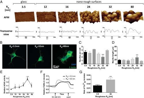
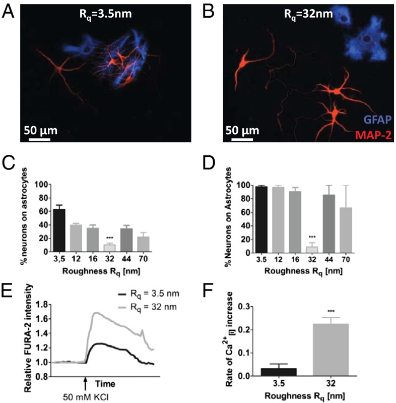
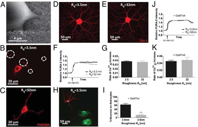
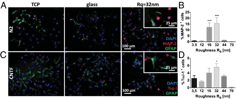
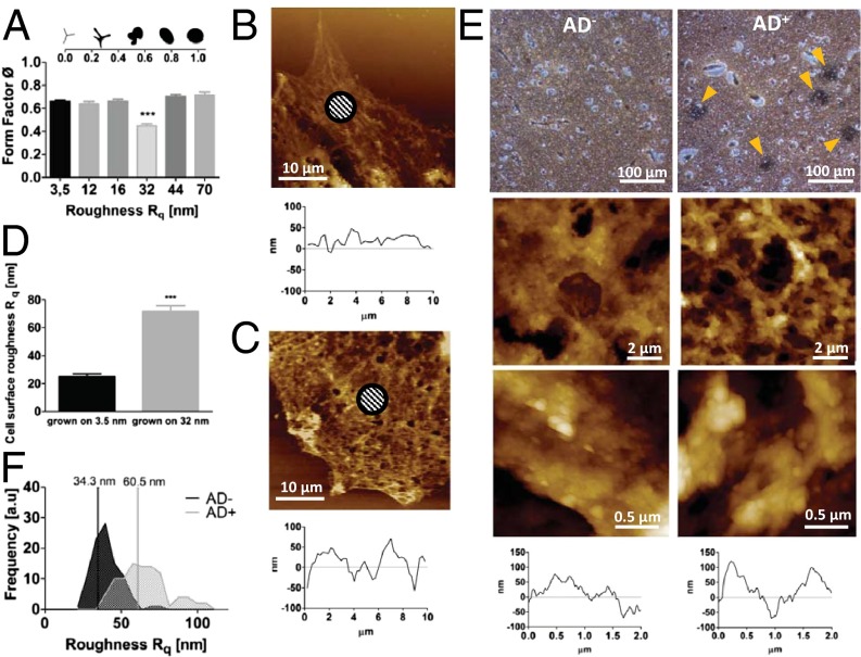
Extracellular soluble signals are known to play a critical role in maintaining neuronal function and homeostasis in the CNS. However, the CNS is also composed of extracellular matrix macromolecules and glia support cells, and the contribution of the physical attributes of these components in maintenance and regulation of neuronal function is not well understood. Because these components possess well-defined topography, we theorize a role for topography in neuronal development and we demonstrate that survival and function of hippocampal neurons and differentiation of telencephalic neural stem cells is modulated by nanoroughness. At roughnesses corresponding to that of healthy astrocytes, hippocampal neurons dissociated and survived independent from astrocytes and showed superior functional traits (increased polarity and calcium flux). Furthermore, telencephalic neural stem cells differentiated into neurons even under exogenous signals that favor astrocytic differentiation. The decoupling of neurons from astrocytes seemed to be triggered by changes to astrocyte apical-surface topography in response to nanoroughness. Blocking signaling through mechanosensing cation channels using GsMTx4 negated the ability of neurons to sense the nanoroughness and promoted decoupling of neurons from astrocytes, thus providing direct evidence for the role of nanotopography in neuron-astrocyte interactions. We extrapolate the role of topography to neurodegenerative conditions and show that regions of amyloid plaque buildup in brain tissue of Alzheimer's patients are accompanied by detrimental changes in tissue roughness. These findings suggest a role for astrocyte and ECM-induced topographical changes in neuronal pathologies and provide new insights for developing therapeutic targets and engineering of neural biomaterials.

Keywords: FAM38A; Piezo-1; mechanotransduction; polarization; stretch-activated channels.


Conflict of interest statement

The authors declare no conflict of interest.

Figures

Fig. 1.
Fig. 1.
Morphological and functional traits in PC-12 cells on nanorough substrates. (A) AFM of SNP modified substrates with the corresponding surface roughness Rq. (B) Morphology of PC-12 cells on Rq = 3.5 nm, Rq = 32 nm, and Rq = 80 nm visualized by staining for F-actin. Impact of nanoroughness on PC-12 polarization as assessed by determining (C) number of neurites per cell and (D) neurite length. (E) Influence of nanoroughness on AChE activity. Calcium-sensitive FURA-2 imaging of differentiated PC-12 cells on smooth glass substrates and surfaces with an Rq of 32 nm: (F) change in intracellular calcium levels as assessed by FURA-2 intensity and (G) rate of depolarization as determined by the slope of the depolarization portion of the curve (immediately after addition of KCl). Statistical significance: *P < 0.05; **P < 0.01; ***P < 0.001.
Fig. 2.
Fig. 2.
Morphology and function of rat hippocampal neurons and astrocytes are influenced by substrate roughnesses. Neuron–astrocyte interaction on (A) smooth glass substrate and (B) substrate of Rq = 32 nm. Astrocytes were visualized using antibody against GFAP (blue) and neurons were visualized using antibody against MAP-2 (red). Quantification of neuron–astrocyte association in (C) short-term cultures (5 d) and (D) long-term cultures (6 wk). Calcium-sensitive FURA-2 imaging in hippocampal neurons on smooth glass substrates and surfaces with Rq of 32 nm: (E) change in intracellular calcium level as assessed by FURA-2 intensity and (F) rate of depolarization as determined by the slope of the depolarization portion of the curve (immediately after addition of KCl). Statistical significance: ***P < 0.001.
Fig. 3.
Fig. 3.
Piezo-1 is necessary for sensing nanotopography. (A) Representative scanning electron micrograph of PC-12 cells grown on nanorough surface (shown Rq = 40 nm). PC-12 stained using anti-FAM38A, an antibody for Piezo-1 mechanosensitive ion channel on Rq = 3.5 nm (B) and Rq = 32 nm (C). On smooth surfaces, FAM38A staining is pronounced at neurite branch points (denoted by circles) (B), whereas on nanorough surfaces FAM38A staining is uniform along all neurite processes (C). Rat dorsal root ganglia morphology (D and E) and function (F and G) on glass and Rq of 32 nm. Inhibition of FAM38A with GsMTx4 (5 µM) results in decoupling of hippocampal neurons from astrocytes on smooth glass substrates (H and I). The FURA-2 intensity profile (J) and rate of calcium influx (K) in hippocampal neurons upon depolarization with KCl on smooth glass substrate and Rq = 32 nm is identical upon inhibition of FAM38A. Statistical significance: ***P < 0.001.
Fig. 4.
Fig. 4.
Nanoroughness mediates rat telencephalic NSC lineage commitment and differentiation. NSC differentiation on TCPS glass, and substrates with Rq of 32 nm. (A) Spontaneous differentiation in N2 media in absence of any exogenous soluble signals (blue, DAPI nuclear stain; red, MAP-2; green, GFAP astrocyte marker), (B) quantification of Map-2+ NSC in the absence of growth factors, (C) differentiation in the presence of CNTF (blue, DAPI nuclear stain; red, Tuj-1; green, GFAP astrocyte marker), and (D) quantification of Tuj-1+ NSC when exposed to CNTF. Statistical significance: *P < 0.05; ***P < 0.001.
Fig. 5.
Fig. 5.
Nanoroughness alters physical attributes of astrocytes. Dependence of astrocyte form factor on substrate nanoroughness. (A) Decrease in form factor on 32-nm Rq surfaces is consistent with a more motile phenotype (Inset). Morphological changes to astrocyte cell surface on nanorough surfaces: AFM images of astrocytes grown on glass (B) and on Rq = 32 nm (C) and the corresponding transverse line scans and changes to topography of astrocyte surface on 32-nm surfaces (D). Shaded areas show representative areas for Rq calculations. Amyloid-β plaques are associated with topographical changes to brain tissue. Paraffin-embedded human brain slices stained with Bielschowsky’s silver stain (E). (Left) Healthy human (AD). (Right) Patient diagnosed with Alzheimer’s (AD+) revealing amyloid-β plaques indicated by yellow arrowheads. (Middle and Bottom) Tapping mode AFM scans (10 × 10 μm) of one of the representative areas above, higher-magnification (2 × 2 μm) scan of the same area, and transverse views of the corresponding 2- × 2-µm image above. (F) Histograms of Rq values of healthy brain tissue and amyloid-β plaques in Alzheimer’s patients showing a general shift of tissue roughness to higher Rqs and an increased heterogeneity in roughness in AD+ brain slices. Rq values were calculated using a 700- × 700-nm scan area.

References

    1. Álvarez-Palazuelos LE, et al. Regulation of neural stem cell in the human SVZ by trophic and morphogenic factors. Curr Signal Transduct Ther. 2011;6(3):320–326. - PMC - PubMed
    1. Steindler DA, Pincus DW. Stem cells and neuropoiesis in the adult human brain. Lancet. 2002;359(9311):1047–1054. - PubMed
    1. Dessaud E, et al. Interpretation of the sonic hedgehog morphogen gradient by a temporal adaptation mechanism. Nature. 2007;450(7170):717–720. - PubMed
    1. Deverman BE, Patterson PH. Cytokines and CNS development. Neuron. 2009;64(1):61–78. - PubMed
    1. Rowitch DH, Kriegstein AR. Developmental genetics of vertebrate glial-cell specification. Nature. 2010;468(7321):214–222. - PubMed

Publication types