Review
. 2014 Dec 5;369(1657):20130537.
doi: 10.1098/rstb.2013.0537.

Primitive endoderm differentiation: from specification to epithelium formation

Affiliations
Review

Primitive endoderm differentiation: from specification to epithelium formation

Stéphanie Hermitte et al. Philos Trans R Soc Lond B Biol Sci. .

Abstract

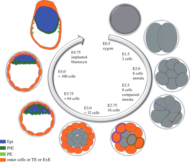
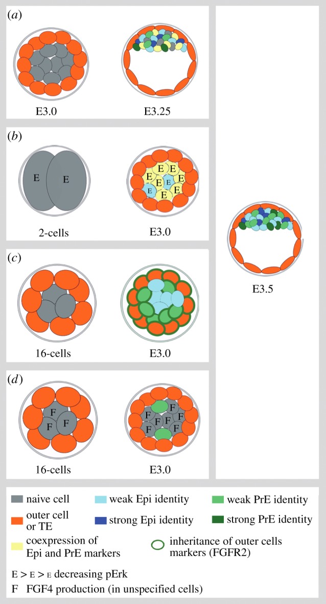
In amniotes, primitive endoderm (PrE) plays important roles not only for nutrient support but also as an inductive tissue required for embryo patterning. PrE is an epithelial monolayer that is visible shortly before embryo implantation and is one of the first three cell lineages produced by the embryo. We review here the molecular mechanisms that have been uncovered during the past 10 years on PrE and epiblast cell lineage specification within the inner cell mass of the blastocyst and on their subsequent steps of differentiation.

Keywords: blastocyst; cell differentiation; cell polarization; cell sorting; epithelium; lineage specification.


Figures

Figure 1.
Figure 1.
Schematic of cell lineages differentiation during mouse preimplantation. After fertilization, the embryo reaches the 8-cell stage and undergoes compaction. During the next two rounds of division, cells divide either symmetrically (purple arrow and cell) or asymmetrically (brown arrow and cell) to give rise to two outer cells or to one outer cell and one inner cell, respectively. Cells can also be internalized. From E3.0 to E3.25 inner cells start to adopt Epi or PrE signatures. At E3.75, a high majority of cells have specified into Epi or PrE. At E4.0, Epi and PrE cells have sorted to form two distinct tissues and have lost their plasticity. By E4.75, PrE cells have formed an epithelium and started to produce parietal endoderm cells that migrate along the trophectoderm. ExE, extraembryonic ectoderm (trophectoderm derivative); PE, parietal endoderm; TE, trophectoderm.
Figure 2.
Figure 2.
Proposed models for the initiation of the salt and pepper pattern. (a) Stochasticity. Before E3.25, inner cells express some Epi and PrE markers randomly. From E3.25, cells progressively acquire either an Epi or a PrE signature, owing to cell-to-cell stochastic expression reinforced by cell interactions. By E3.75, the two lineages are distinct [23]. (b) Nanog expression gain through pErk decrease. Cells express high levels of active Erk at the two cells stage, owing to high levels of FGF4. The transcription of Fgf4 diminishes subsequently. From the 8-cell stage, Nanog and Gata6 are coexpressed in all the cells. Around E3.0–E3.25, the FGF4 protein levels are heterogeneous leading to a lower Erk activity in some cells. As a consequence, these cells promote Nanog expression, which in turn re-induces FGF4 expression at high levels. The neighbouring cells receive the FGF4 message and differentiate into PrE. The specification of the two lineages is asynchronous and leads to a progressive cell-to-cell identity acquisition [20]. (c) Inherent differences between In1 and In2 cells. At the 32-cell stage (E3.0), In2 cells inherit FGF2 from their mothers. As a consequence, In2 cells preferentially become PrE cells, whereas In1 cells favour an Epi identity [52]. In1 cells could acquire an Epi identity as soon as they are internalized or later, as a default state. (d) Inside cell accumulation. Inner cells (In1 and In2) produce FGF4. The accumulation of FGF4-expressing cells leads to high concentration of secreted FGF4 that is sufficient to induce PrE differentiation in some cells [53]. Epi identity could be acquired as an induced or default state.

References

    1. Rossant J, Tam PP. 2009. Blastocyst lineage formation, early embryonic asymmetries and axis patterning in the mouse. Development 136, 701–713. (10.1242/dev.017178) - DOI - PubMed
    1. Kwon GS, Viotti M, Hadjantonakis AK. 2008. The endoderm of the mouse embryo arises by dynamic widespread intercalation of embryonic and extraembryonic lineages. Dev. Cell 15, 509–520. (10.1016/j.devcel.2008.07.017) - DOI - PMC - PubMed
    1. Yamanaka Y, Lanner F, Rossant J. 2010. FGF signal-dependent segregation of primitive endoderm and epiblast in the mouse blastocyst. Development 137, 715–724. (10.1242/dev.043471) - DOI - PubMed
    1. McDole K, Xiong Y, Iglesias PA, Zheng Y. 2011. Lineage mapping the pre-implantation mouse embryo by two-photon microscopy, new insights into the segregation of cell fates. Dev. Biol. 355, 239–249. (10.1016/j.ydbio.2011.04.024) - DOI - PMC - PubMed
    1. Morris SA, Teo RT, Li H, Robson P, Glover DM, Zernicka-Goetz M. 2010. Origin and formation of the first two distinct cell types of the inner cell mass in the mouse embryo. Proc. Natl Acad. Sci. USA 107, 6364–6369. (10.1073/pnas.0915063107) - DOI - PMC - PubMed

Publication types

MeSH terms

LinkOut - more resources