Review
. 2014 Dec 5;369(1657):20130538.
doi: 10.1098/rstb.2013.0538.

Developmental plasticity, cell fate specification and morphogenesis in the early mouse embryo

Affiliations
Review

Developmental plasticity, cell fate specification and morphogenesis in the early mouse embryo

Ivan Bedzhov et al. Philos Trans R Soc Lond B Biol Sci. .

Erratum in

Abstract

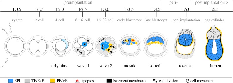
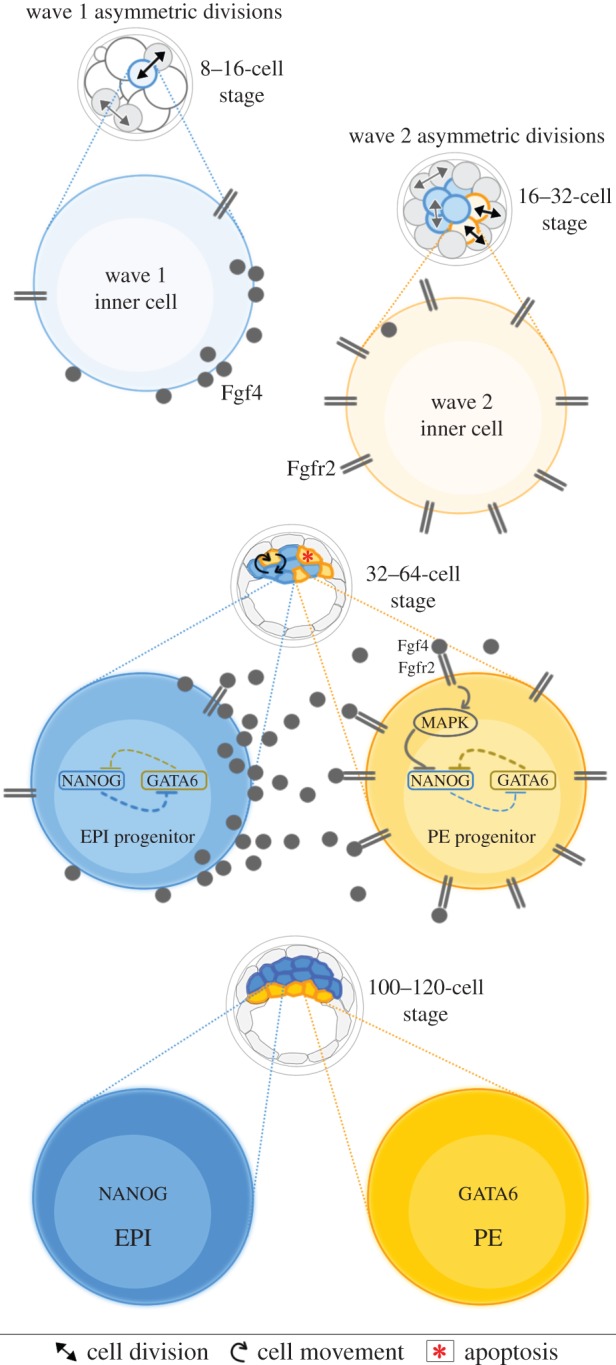
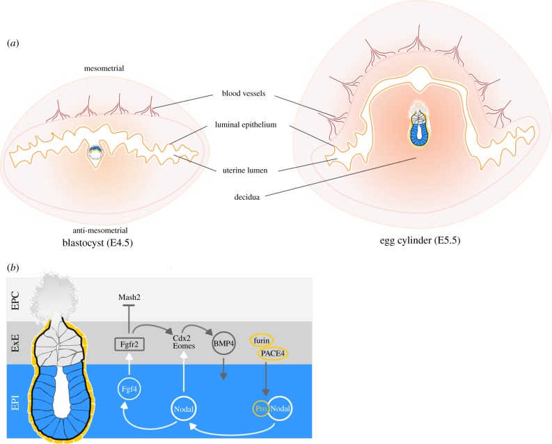
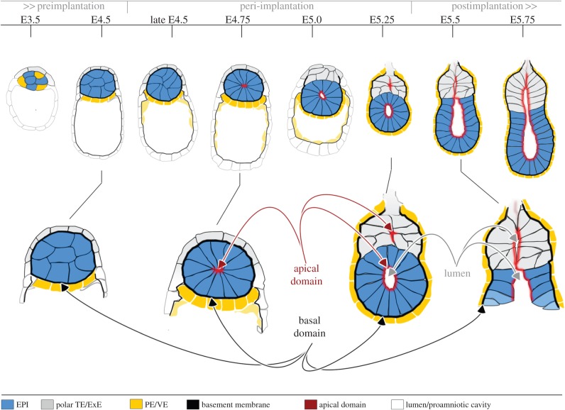
A critical point in mammalian development is when the early embryo implants into its mother's uterus. This event has historically been difficult to study due to the fact that it occurs within the maternal tissue and therefore is hidden from view. In this review, we discuss how the mouse embryo is prepared for implantation and the molecular mechanisms involved in directing and coordinating this crucial event. Prior to implantation, the cells of the embryo are specified as precursors of future embryonic and extra-embryonic lineages. These preimplantation cell fate decisions rely on a combination of factors including cell polarity, position and cell-cell signalling and are influenced by the heterogeneity between early embryo cells. At the point of implantation, signalling events between the embryo and mother, and between the embryonic and extraembryonic compartments of the embryo itself, orchestrate a total reorganization of the embryo, coupled with a burst of cell proliferation. New developments in embryo culture and imaging techniques have recently revealed the growth and morphogenesis of the embryo at the time of implantation, leading to a new model for the blastocyst to egg cylinder transition. In this model, pluripotent cells that will give rise to the fetus self-organize into a polarized three-dimensional rosette-like structure that initiates egg cylinder formation.

Keywords: cell fate; differentiation; embryo; morphogenesis; pluripotency.


Figures

Figure 1.
Figure 1.
Overview of early mouse development. Embryonic and extraembryonic cells are specified in the preimplantation embryo by two cell fate decisions. In the first cell fate decision, waves of cell divisions create inside and outside cells. Outside cells give rise to extraembryonic trophectoderm (TE), while inside cells form the pluripotent inner cell mass (ICM). In the second cell fate decision, cells of the ICM are segregated into the extraembryonic PE and the pluripotent epiblast (EPI) that will later give rise to all tissues of the body. These fate decisions are influenced but not determined by heterogeneity between individual cells within the embryo that is established by the 4-cell stage (shown by different shading of cells). At E4.5, the embryo initiates implantation and over the next 24 h invades the maternal tissues, rapidly proliferates and transforms into an egg cylinder. This new form serves as a foundation for EPI patterning, laying down the body axis and establishment of the germ layers. ExE, extraembryonic ectoderm; PE, primitive endoderm; VE, visceral endoderm.
Figure 2.
Figure 2.
Specification of the TE and ICM. The compacted morula is the earliest point where blastomeres have differential spatial positioning. Blastomeres on the inside of the embryo encounter symmetric cell–cell contact and give rise to the pluripotent ICM. Outside cells have asymmetric cell–cell contact and form the extraembryonic TE. The asymmetry in cell–cell contact leads to accumulation of polarity proteins such as Par6 and aPKC at the apical domain. Par6 and aPKC activity inhibits activity of Amot and the Hippo pathway kinases Lats1/2. As a result, Yap and Taz are de-repressed, Tead4 transcriptional activity is switched on and Cdx2 expression and the TE cell fate programme are activated. In inside cells, symmetric cell–cell contact prevents establishment of an apical domain. Amot is active and is likely tethered to AJs through a putative Nf2–α-catenin–β-catenin–E-cadherin complex. Amot sequesters Yap/Taz to the cytoplasm as well as activating the Hippo pathway proteins Lats1/2. Lats1/2 can inhibit Yap/Taz activity via the canonical Hippo pathway and can also activate Amot in a positive feedback loop. In the absence of Yap/Taz in the nucleus, Tead4 activity is switched off, Oct4 expression is promoted and the default pluripotent programme is expressed. Therefore, positional and polarity cues in these two populations lead to changes in gene regulation and differentiation into their respective lineages.
Figure 3.
Figure 3.
Specification of the EPI and PE. ICM cells internalized in the first wave of asymmetric cell divisions upregulate Fgf4, while cells internalized in the second wave express higher levels of Fgfr2, possibly by inheriting this transcription factor from their outside 16-cell stage mother cells. Fgf signalling in wave two cells inhibits the repression of Gata6 expression by Nanog, biasing these cells towards the PE lineage.
Figure 4.
Figure 4.
Implantation and signalling during egg cylinder formation. (a) After hatching, blastocyst adheres to the LE and invades the stroma at the antimesometrial site of the uterus. In response, the stromal cells differentiate into decidual cells that regulate trophoblast invasion, enable nutrients and gas exchange and ensure fetomaternal immune tolerance. In the next 24 h, the decidua rapidly proliferates to support embryo development into the egg cylinder and beyond. (b) The EPI cells secrete Fgf4 ligand that binds to Fgfr2 in the ExE in a paracrine manner. Active Fgfr2 signalling promotes Cdx2 and Eomes expression and inhibits genes associated with TS cell differentiation such as Mash2. Nodal produced in the EPI promotes ExE maintenance directly by activating core TS cell genes and indirectly by sustaining Fgf4 expression. In turn, ExE potentiates Nodal activity by secreting furin and PACE4 proteases that cleave out the Nodal propeptide to generate a mature ligand with higher activity.
Figure 5.
Figure 5.
Model of peri-implantation morphogenesis. Following preimplantation lineage segregation (E3.5–E4.5), the extraembryonic lineages start secreting ECM proteins that assemble a basal membrane that wraps around the EPI and provides polarization cues through integrin receptors. During the peri-implantation period (late E4.5–E5.0), the pluripotent EPI cells establish apical–basal polarity, change shape and constrict apically while clustering to form a rosette. A central lumen emerges in the centre of the rosette through hollowing of apical membranes by charge repulsion (E5.0–E5.25). As the egg cylinder elongates the lumen enlarges and incorporates intramembranous spaces of the proximal ExE to form the mature pro-amniotic cavity (E5.5–E5.75).

References

    1. Johnson MH, Ziomek CA. 1981. The foundation of two distinct cell lineages within the mouse morula. Cell 24, 71–80. (10.1016/0092-8674(81)90502-X) - DOI - PubMed
    1. Morris SA, Teo RTY, Li H, Robson P, Glover DM, Zernicka-Goetz M. 2010. Origin and formation of the first two distinct cell types of the inner cell mass in the mouse embryo. Proc. Natl Acad. Sci. USA 107, 6364–6369. (10.1073/pnas.0915063107) - DOI - PMC - PubMed
    1. Fleming TP. 1987. A quantitative analysis of cell allocation to trophectoderm and inner cell mass in the mouse blastocyst. Dev. Biol. 119, 520–531. (10.1016/0012-1606(87)90055-8) - DOI - PubMed
    1. Suwińska A, Czołowska R, Ożdżeński W, Tarkowski AK. 2008. Blastomeres of the mouse embryo lose totipotency after the fifth cleavage division: expression of Cdx2 and Oct4 and developmental potential of inner and outer blastomeres of 16- and 32-cell embryos. Dev. Biol. 322, 133–144. (10.1016/j.ydbio.2008.07.019) - DOI - PubMed
    1. Tarkowski AK. 1959. Experiments on the development of isolated blastomers of mouse eggs. Nature 184, 1286–1287. (10.1038/1841286a0) - DOI - PubMed

Publication types

LinkOut - more resources