Review
. 2014 Oct 28;6(10):741-55.
doi: 10.4329/wjr.v6.i10.741.

2-deoxy-2-((18)F)fluoro-D-glucose positron emission tomography/computed tomography imaging in paediatric oncology

Affiliations
Review

2-deoxy-2-((18)F)fluoro-D-glucose positron emission tomography/computed tomography imaging in paediatric oncology

John Freebody et al. World J Radiol. .

Abstract

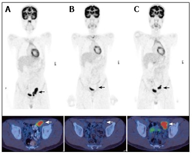
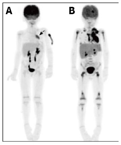
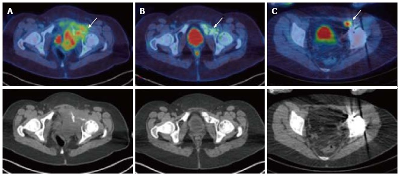
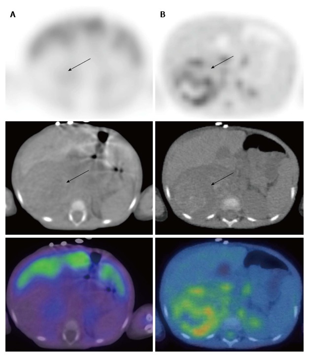
Positron emission tomography (PET) is a minimally invasive technique which has been well validated for the diagnosis, staging, monitoring of response to therapy, and disease surveillance of adult oncology patients. Traditionally the value of PET and PET/computed tomography (CT) hybrid imaging has been less clearly defined for paediatric oncology. However recent evidence has emerged regarding the diagnostic utility of these modalities, and they are becoming increasingly important tools in the evaluation and monitoring of children with known or suspected malignant disease. Important indications for 2-deoxy-2-((18)F)fluoro-D-glucose (FDG) PET in paediatric oncology include lymphoma, brain tumours, sarcoma, neuroblastoma, Langerhans cell histiocytosis, urogenital tumours and neurofibromatosis type I. This article aims to review current evidence for the use of FDG PET and PET/CT in these indications. Attention will also be given to technical and logistical issues, the description of common imaging pitfalls, and dosimetric concerns as they relate to paediatric oncology.

Keywords: Computed tomography; Dosimetry; Fluorodeoxyglucose F18; Oncology; Paediatrics; Positron emission tomography; Technical issues.


Figures

Figure 1
Figure 1
A 13 year-old male with nodular sclerosing Hodgkin’s disease. PET/CT at staging (A) demonstrated disease in the left external iliac region. After completion of chemotherapy 4 mo later (B) there was a complete metabolic response with no activity in the residual lymph node mass. PET/CT performed 6 mo later (C) for surveillance demonstrated a recurrence at the same site. PET/CT: Positron emission tomography/computed tomography.
Figure 2
Figure 2
A 5-year-old male patient with relapsed T-cell lymphoblastic lymphoma. Restaging PET at the time of relapse 1 year after initial therapy demonstrated an FDG-avid mediastinal mass (A, arrow). Repeat PET performed following 1 cycle of FLAG-Ida chemotherapy demonstrated no response to treatment and progression of disease to stage IV with extensive bone marrow involvement (B). PET: Positron emission tomography; FDG: 2-deoxy-2-(18F)fluoro-D-glucose.
Figure 3
Figure 3
A 17-year-old female with Ewing’s sarcoma involving the left superior pubic ramus. Staging PET/CT showed extensive disease (A, arrow) with bone destruction and a large FDG-avid pelvic mass. Following chemotherapy, at 5 mo after diagnosis, there was an excellent metabolic response with minimal residual FDG uptake (B, arrow). Patient underwent surgical resection and extracorporeal radiotherapy to the bone. Sixteen months after completion of treatment, surveillance PET/CT demonstrated recurrence in a left external iliac lymph node (C) which was not detectable on the CT or MRI due to marked metal artefact. PET/CT: Positron emission tomography/computed tomography; FDG: 2-deoxy-2-(18F)fluoro-D-glucose; MRI: Magnetic resonance imaging.
Figure 4
Figure 4
Neuroblastoma in a 2-year-old female. 123I-metaiodobenzylguanidine (MIBG) single positron emission tomography/computed tomography (SPECT/CT) images (A) demonstrated a large right suprarenal mass displacing the organs which was not MIBG-avid (arrows). FDG PET/CT (B) showed moderate heterogenous metabolic activity within the mass. PET/CT: Positron emission tomography/computed tomography; FDG: 2-deoxy-2-(18F)fluoro-D-glucose.
Figure 5
Figure 5
A 7-year-old male with Langerhans cell histiocytosis. Skeletal survey demonstrated an isolated left femoral lesion, confirmed on PET/CT (A, arrow). Additional lesions (arrows) in the right 6th rib posteriorly (B) and in the skull (C) were also identified on the PET/CT scan. Top panel: fused PET/CT images, bottom panel: low dose CT component of the scan. PET/CT: Positron emission tomography/computed tomography.
Figure 6
Figure 6
An extreme case of widespread brown fat activity in a 16-year-old female with treated Hodgkin’s disease.
Figure 7
Figure 7
Normal physiological variants: various patterns of thymic uptake (A-F, arrow), tonsillar uptake (B, arrowhead), cardiac uptake (D, arrowhead) and laryngeal uptake (F, arrowhead).
Figure 8
Figure 8
Examples of abnormal thymic uptake due to malignant disease (arrows).
Figure 9
Figure 9
Normal 2-deoxy-2-(18F)fluoro-D-glucose positron emission tomography scan of an 8-year-old female showing physiological growth plate uptake (arrow).
Figure 10
Figure 10
Non–ossifying fibroma (arrow) in the left distal femur of a 17-year-old male patient with Langerhans cell histiocytosis. FDG PET (A) showing avid uptake, CT (B) showing typical benign radiological features. PET: Positron emission tomography; CT: Computed tomography; FDG: 2-deoxy-2-(18F)fluoro-D-glucose.
Figure 11
Figure 11
Effective dose vs patient weight for 2-deoxy-2-(18F)fluoro-D-glucose (European Association of Nuclear Medicine and North American Consensus Guidelines). Image courtesy of Bruce McBride, Chief Physicist, Department of Nuclear Medicine and PET, The Prince of Wales and Sydney Children’s Hospitals. PET: Positron emission tomography; EANM: European Association of Nuclear Medicine.

Similar articles

Cited by

References

    1. Gatta G, Zigon G, Capocaccia R, Coebergh JW, Desandes E, Kaatsch P, Pastore G, Peris-Bonet R, Stiller CA; EUROCARE Working Group. Survival of European children and young adults with cancer diagnosed 1995-2002. Eur J Cancer. 2009;45:992–1005. - PubMed
    1. Ouyang J, Li Q, El Fakhri G. Magnetic resonance-based motion correction for positron emission tomography imaging. Semin Nucl Med. 2013;43:60–67. - PMC - PubMed
    1. Juweid ME, Cheson BD. Positron-emission tomography and assessment of cancer therapy. N Engl J Med. 2006;354:496–507. - PubMed
    1. Storto G, Nicolai E, Salvatore M. [18F]FDG-PET-CT for early monitoring of tumor response: when and why. Q J Nucl Med Mol Imaging. 2009;53:167–180. - PubMed
    1. Wegner EA, Barrington SF, Kingston JE, Robinson RO, Ferner RE, Taj M, Smith MA, O’Doherty MJ. The impact of PET scanning on management of paediatric oncology patients. Eur J Nucl Med Mol Imaging. 2005;32:23–30. - PubMed