

Recent advances in imaging technologies in dentistry
- PMID: 25349663
- PMCID: PMC4209425
- DOI: 10.4329/wjr.v6.i10.794
Recent advances in imaging technologies in dentistry
Abstract
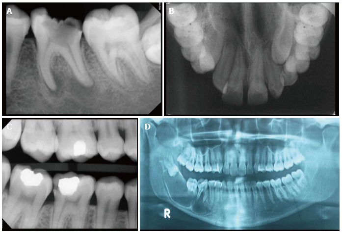
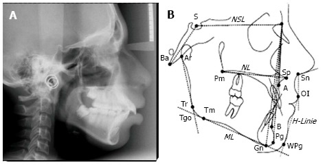
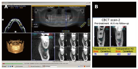
Dentistry has witnessed tremendous advances in all its branches over the past three decades. With these advances, the need for more precise diagnostic tools, specially imaging methods, have become mandatory. From the simple intra-oral periapical X-rays, advanced imaging techniques like computed tomography, cone beam computed tomography, magnetic resonance imaging and ultrasound have also found place in modern dentistry. Changing from analogue to digital radiography has not only made the process simpler and faster but also made image storage, manipulation (brightness/contrast, image cropping, etc.) and retrieval easier. The three-dimensional imaging has made the complex cranio-facial structures more accessible for examination and early and accurate diagnosis of deep seated lesions. This paper is to review current advances in imaging technology and their uses in different disciplines of dentistry.
Keywords: Cephalogram; Dental X-rays; Dental cone beam computed tomography; Intraoral X-rays; Panoramic radiograph.
Figures
References
-
- Nair MK, Nair UP. Digital and advanced imaging in endodontics: a review. J Endod. 2007;33:1–6. - PubMed
-
- Mouyen F, Benz C, Sonnabend E, Lodter JP. Presentation and physical evaluation of RadioVisioGraphy. Oral Surg Oral Med Oral Pathol. 1989;68:238–242. - PubMed
-
- Langland OE, Langlais RP, Preece JW. Principles of dental imaging. 2nd ed. Philadelphia: Lippincott Williams & Wilkins; 2002. p. 285.
-
- Frederiksen NL. Health Physics. In: Pharoah MJ, White SC, editors. Oral Radiology Principles and Interpretation. 4th ed. Mosby: St. Louis; 2000. p. 53.
-
- Visser H, Rödig T, Hermann KP. Dose reduction by direct-digital cephalometric radiography. Angle Orthod. 2001;71:159–163. - PubMed
Publication types
LinkOut - more resources
Full Text Sources
Other Literature Sources
Miscellaneous
