

Concurrent intracranial and spinal subdural hematoma in a teenage athlete: a case report of this rare entity
- PMID: 25349764
- PMCID: PMC4198776
- DOI: 10.1155/2014/143408
Concurrent intracranial and spinal subdural hematoma in a teenage athlete: a case report of this rare entity
Abstract
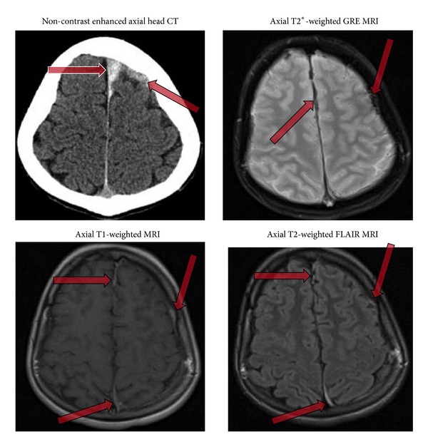
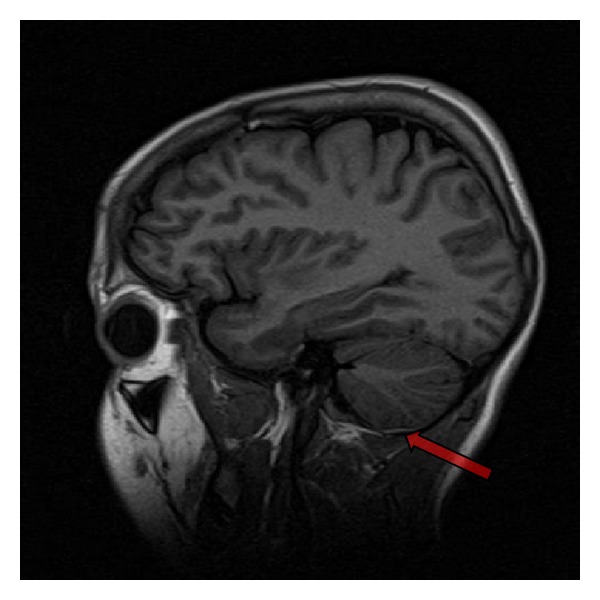
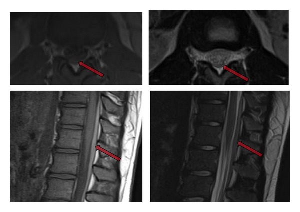
A 15-year-old male high school football player presented with episodes of headache and complete body stiffness, especially in the arms, lower back, and thighs, immediately following a football game. This was accompanied by severe nausea and vomiting for several days. Viral meningitis was suspected by the primary clinician, and treatment with corticosteroids was initiated. Over the next several weeks, there was gradual symptom improvement and the patient returned to his baseline clinical status. The patient experienced a severe recurrence of the previous myriad of symptoms following a subsequent football game, without an obvious isolated traumatic episode. In addition, he experienced a new left sided headache, fatigue, and difficulty ambulating. He was admitted and an extensive workup was performed. CT and MRI of the head revealed concurrent intracranial and spinal subdural hematomas (SDH). Clinical workup did not reveal any evidence of coagulopathy or predisposing vascular lesions. Spinal SDH is an uncommon condition whose concurrence with intracranial SDH is an even greater clinical rarity. We suggest that our case represents an acute on chronic intracranial SDH with rebleeding, membrane rupture, and symptomatic redistribution of hematoma to the spinal subdural space.
Figures
References
-
- Kreppel D, Antoniadis G, Seeling W. Spinal hematoma: a literature survey with meta-analysis of 613 patients. Neurosurgical Review. 2003;26(1):1–49. - PubMed
-
- Kokubo R, Kim K, Mishina M, et al. Prospective assessment of concomitant lumbar and chronic subdural hematoma: is migration from the intracranial space involved in their manifestation? Journal of Neurosurgery: Spine. 2014;20(2):157–163. - PubMed
-
- Khosla VK, Kak VK, Mathuriya SN. Chronic spinal subdural hematomas: report of two cases. Journal of Neurosurgery. 1985;63(4):636–639. - PubMed
-
- Cronberg S, Wallmark E, Soderberg I. Effect on platelet aggregation of oral administration of 10 non-steroidal analgesics to humans. Scandinavian Journal of Haematology. 1984;33(2):155–159. - PubMed
LinkOut - more resources
Full Text Sources
Other Literature Sources
