. 2015 Aug;103(6):1217-1227.
doi: 10.1002/jbm.b.33299. Epub 2014 Oct 28.

Equine model for soft-tissue regeneration

Affiliations

Equine model for soft-tissue regeneration

E Bellas et al. J Biomed Mater Res B Appl Biomater. 2015 Aug.

Abstract

Soft-tissue regeneration methods currently yield suboptimal clinical outcomes due to loss of tissue volume and a lack of functional tissue regeneration. Grafted tissues and natural biomaterials often degrade or resorb too quickly, while most synthetic materials do not degrade. In previous research we demonstrated that soft-tissue regeneration can be supported using silk porous biomaterials for at least 18 months in vivo in a rodent model. In the present study, we scaled the system to a survival study using a large animal model and demonstrated the feasibility of these biomaterials for soft-tissue regeneration in adult horses. Both slow and rapidly degrading silk matrices were evaluated in subcutaneous pocket and intramuscular defect depots. We showed that we can effectively employ an equine model over 6 months to simultaneously evaluate many different implants, reducing the number of animals needed. Furthermore, we were able to tailor matrix degradation by varying the initial format of the implanted silk. Finally, we demonstrate ultrasound imaging of implants to be an effective means for tracking tissue regeneration and implant degradation.

Keywords: animal model; in vivo test; mesenchymal stem cell; scaffold; silk.


Figures

Fig. 1
Fig. 1
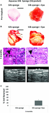
Aqueous silk sponge implants in subcutaneous pocket model. (A) Aqueous silk sponges prior to implantation, left- aqueous sponge alone, right- aqueous silk sponge seeded with lipo. Scale bar 5mm. (B) Implants after harvest at 3 months. Top row - macroscopic appearance of implants, scale bar 5mm. Implants have a natural tissue-like appearance and silk structure is not grossly detectable. Middle row - H&E sections of aqueous silk sponge (black arrowhead) no longer has pore structure as it degrades, scale bar 100μm, inset 200μm. Bottom row- representative ultrasound images of implant site with implant encircled in dotted line. (C) The aqueous silk sponge alone had degraded into small fractions after 3 months and therefore volume measurements were not possible, however the aqueous silk sponge with lipo retained 37 ±10 % of its volume.
Fig. 2
Fig. 2
Solvent silk sponge implants in subcutaneous pocket model. (A) Solvent silk sponges prior to implantation, left- solvent sponge alone, right- solvent silk sponge seeded with lipo. Scale bar 5mm. (B) Implants after harvest at 6 months. Top row- macroscopic appearance of implants, scale bar 5mm. Implants retain a porous spongy appearance and silk structure is clearly visible in solvent sponge alone, whereas the solvent sponge with lipo has a more tissue-like appearance. Middle row- H&E sections of solvent silk sponge (black arrowhead) maintained pore structure, scale bar 100μm, inset 200μm. Bottom row- representative ultrasound images of implant site with implant encircled in dotted line. (C) Both solvent silk sponge groups maintained close to their initial volumes.
Fig. 3
Fig. 3
Subcutaneous silk foam implant volume increases with implant time. (A) Silk foams prior to implantation, left- foam alone, right- foam seeded with lipo. Scale bar 5mm. (B) Silk foam appearance after 1 month. Top row- macroscopic appearance of implants, scale bar 5mm. Silk foam implants alone still maintain their foam structure appearance. When foams are seeded with lipo the result is a more natural tissue-like appearance and silk structure is not as detectable. Scale bar 5mm. Middle row- Silk foam (black arrowhead) clearly visible in H&E sections, scale bar 100μm, inset 200μm. Bottom row- representative ultrasound images of implant site with implant encircled in dotted line. (C) Silk foam appearance after 3 months. Top row- macroscopic appearance of implants, scale bar 5mm. Silk foam implants alone maintain less of their foam structure appearance than at 1 month. When foams are seeded with lipo the result is a more natural tissue-like appearance and silk structure is even less detectable. Scale bar 5mm. Middle row- Silk foam (black arrowhead) clearly visible in H&E sections, scale bar 100μm, inset 200μm. Bottom row- representative ultrasound images of implant site with implant encircled in dotted line. (D) At 1 month only about half of the initial volume remained, yet at 3 months the initial volume was regained. Volume was unaffected by addition of lipo.
Fig. 4
Fig. 4
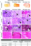
Intramuscular defect repair with silk sponges. (A) Sponge appearance prior to implantation (both aqueous and solvent silk sponges had same macroscopic appearance and therefore only aqueous sponges are shown for simplicity). Scale bar 20mm. (B) Aqueous silk sponge after intramuscular implantation at 1 month. H&E images of aqueous silk sponges at 1 month. The pore structure of silk sponges (black arrowhead) was no longer intact. When seeded with lipo, matrix-dense (white arrow) areas were seen. No muscle was seen. Scale bar 100μm, inset 200μm. (C) H&E images of aqueous silk sponges at 3 months. Fewer cells and more matrix were present than at 1 month. Scale bar 100μm, inset 200μm. (D) Intramuscular implants at 6 months. H&E images of solvent silk sponge groups. Pore structure of silk sponges clearly visible in all groups (black arrowhead). No muscle regeneration was seen within the boundaries of the silk implants. When silk sponges were seeded with lipo, there was a band (~100 μm thick) of muscle present immediately adjacent to the implant (white arrow). (E) Intramuscular defect sites at 6 months without silk sponges. Defect alone (left image) had a scar like appearance, while UMSCs injected at the defect site (middle image) yielded some muscle regeneration (bottom bracket) and some scar tissue (top bracket). Improved muscle regeneration was visible in the lipo defect group. Scale bar 100μm, inset 200μm. (F) Aqueous silk sponges had begun to degrade at 1 month and 3 months. Solvent based silk sponges had degraded to a similar extent at 6 months as aqueous silk sponges did in 1-3 months.

References

    1. Machingal MA, Corona BT, Walters TJ, Kesireddy V, Koval CN, Dannahower A, et al. A tissue-engineered muscle repair construct for functional restoration of an irrecoverable muscle injury in a murine model. Tissue Eng Part A. 2011;17:2291–303. - PMC - PubMed
    1. Corona BT, Machingal MA, Criswell T, Vadhavkar M, Dannahower AC, Bergman C, et al. Further development of a tissue engineered muscle repair construct in vitro for enhanced functional recovery following implantation in vivo in a murine model of volumetric muscle loss injury. Tissue Eng Part A. 2012;18:1213–28. - PMC - PubMed
    1. Stosich MS, Moioli EK, Wu JK, Lee CH, Rohde C, Yoursef AM, et al. Bioengineering strategies to generate vascularized soft tissue grafts with sustained shape. Methods. 2009;47:116–21. - PMC - PubMed
    1. Patrick C. Tissue engineering strategies for adipose tissue repair. Anat Rec. 2001;366:361–6. - PubMed
    1. De Sousa A. Psychological issues in oral and maxillofacial reconstructive surgery. Br J Oral Maxillofac Surg. 2008;46:661–4. - PubMed

Publication types

LinkOut - more resources