. 2014 Oct 28;9(10):e110693.
doi: 10.1371/journal.pone.0110693. eCollection 2014.

β2-Adrenergic receptor-dependent attenuation of hypoxic pulmonary vasoconstriction prevents progression of pulmonary arterial hypertension in intermittent hypoxic rats

Affiliations

β2-Adrenergic receptor-dependent attenuation of hypoxic pulmonary vasoconstriction prevents progression of pulmonary arterial hypertension in intermittent hypoxic rats

Hisashi Nagai et al. PLoS One. .

Abstract

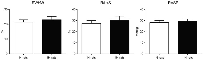
In sleep apnea syndrome (SAS), intermittent hypoxia (IH) induces repeated episodes of hypoxic pulmonary vasoconstriction (HPV) during sleep, which presumably contribute to pulmonary arterial hypertension (PAH). However, the prevalence of PAH was low and severity is mostly mild in SAS patients, and mild or no right ventricular hypertrophy (RVH) was reported in IH-exposed animals. The question then arises as to why PAH is not a universal finding in SAS if repeated hypoxia of sufficient duration causes cycling HPV. In the present study, rats underwent IH at a rate of 3 min cycles of 4-21% O2 for 8 h/d for 6 w. Assessment of diameter changes in small pulmonary arteries in response to acute hypoxia and drugs were performed using synchrotron radiation microangiography on anesthetized rats. In IH-rats, neither PAH nor RVH was observed and HPV was strongly reversed. Nadolol (a hydrophilic β(1, 2)-blocker) augmented the attenuated HPV to almost the same level as that in N-rats, but atenolol (a hydrophilic β1-blocker) had no effect on the HPV in IH. These β-blockers had almost no effect on the HPV in N-rats. Chronic administration of nadolol during 6 weeks of IH exposure induced PAH and RVH in IH-rats, but did not in N-rats. Meanwhile, atenolol had no effect on morphometric and hemodynamic changes in N and IH-rats. Protein expression of the β1-adrenergic receptor (AR) was down-regulated while that of β2AR was preserved in pulmonary arteries of IH-rats. Phosphorylation of p85 (chief component of phosphoinositide 3-kinase (PI3K)), protein kinase B (Akt), and endothelial nitric oxide synthase (eNOS) were abrogated by chronic administration of nadolol in the lung tissue of IH-rats. We conclude that IH-derived activation of β2AR in the pulmonary arteries attenuates the HPV, thereby preventing progression of IH-induced PAH. This protective effect may depend on the β2AR-Gi mediated PI3K/Akt/eNOS signaling pathway.


Conflict of interest statement

Competing Interests: The authors have declared that no competing interests exist.

Figures

Figure 1
Figure 1. Neither right ventricular hypertrophy nor RVSP elevation was observed after 6 weeks of IH exposure.
Heart weight and right ventricular systolic pressure (RVSP) were measured after 6 weeks IH exposure (n = 10). Data are presented as mean ± S.D. RV: right ventricle weight, HW: heart weight, R/L+S (Fulton’s index): right ventricle weight/left ventricle and septum weight.
Figure 2
Figure 2. Selective blockade of peripheral β2AR restored HPV in IH-rats.
(A) Representative microangiogram images showing the branching pattern of small pulmonary arteries during normoxia and in response to hypoxia with or without drugs. Black arrows point to branches of pulmonary arteries that have constricted in response to acute hypoxia. IH has no response to hypoxia, however, significant vasoconstriction is revealed with nadolol. The tungsten wire in the bottom right of each image is a reference of 50 µm diameter. (B) Relationship between vessel size and the magnitude of pulmonary vasoconstriction (% decrease in vessel diameter) in response to acute hypoxia (10% O2 for 10 min) in N-rats and IH-rats with or without β-blocker administration. Data are presented as mean ± S.E.M. *Significant reduction in vessel diameter compared to normoxic condition (** P<0.01). Significant difference between N-rats and IH-rats (†† P<0.01).Significant difference compared to without-drug. (‡‡ P<0.01).
Figure 3
Figure 3. Chronic administration of nadolol induced PAH and RVH in IH-rats without pulmonary arterial hypertrophy.
(A) Hemodynamic and morphometric change after chronic subcutaneous administration of atenolol and nadolol (n = 4 each) during 6weeks of IH exposure. Data are presented as mean ± S.D. *Significant change compared with every other group (* P<0.05, ** P<0.01). (B) Representative images of small pulmonary arteries and assessment of pulmonary arterial hypertrophy by means of medial wall thickness in N and IH-rats with/without chronic administration of atenolol or nadolol (n = 4 each). There were no significant differences in medial wall thickness between each group. Calibration bar = 20 µm. Data are presented as mean ± S.D.
Figure 4
Figure 4. Expression of β1 and β2AR was slightly changed but not significant so in the lung of IH-rats.
Quantitative analysis of β1AR, β2AR, and actin protein expression in whole lung of N-rats and IH-rats (n = 6 each) using Western blot. (A) representative Western blot bands, (B) relative amount of protein. Data are presented as mean ± S.D.
Figure 5
Figure 5. Expression level of β1AR was decreased and β2AR was preserved in the pulmonary arteries of IH-rats.
Quantitative immunohistochemistry of β1 and β2AR was performed in the pulmonary arteries in the diameter range of 50 to 150 µm (n = 6 each). (A) representative images of immunohistochemistry, (B) mean optical density, % area, and expression level score. Quantification of the expression level of the protein was estimated as expression level score (ELS) : ELS = (mean optical density of positively stained area – mean optical density of background area) x percent area of positively stained. *Significant difference between N and IH-rats (** P<0.01).
Figure 6
Figure 6. IH-induced phosphorylation of p85, Akt and eNOS were abrogated by nadolol administration in the lung tissue of IH-rats.
Quantitative analysis of phospho-p85, p85, phospho-Akt, Akt, phospho-eNOS, eNOS, and actin protein expression in whole lung of N-rats and IH-rats (n = 6 each) using Western blot. (A) representative Western blot bands, (B) relative amount of protein. Data are presented as mean ± S.D. *Significant difference between IH-group and each other groups (* P<0.05, ** P<0.01). N: N-rats, NA:N-rats+atenolol, NN: N-rats+nadolol, IH: IH-rats, IA: IH-rats+atenolol, IN: IH-rats+nadolol.

References

    1. Dempsey JA, Veasey SC, Morgan BJ, O'Donnell CP (2010) Pathophysiology of sleep apnea. Physiol Rev 90: 47–112. - PMC - PubMed
    1. Marrone O, Bonsignore MR (2002) Pulmonary haemodynamics in obstructive sleep apnoea. Sleep Med Rev 6: 175–193. - PubMed
    1. Bosc LV, Resta T, Walker B, Kanagy NL (2010) Mechanisms of intermittent hypoxia induced hypertension. J Cell Mol Med 14: 3–17. - PMC - PubMed
    1. Sylvester JT, Shimoda LA, Aaronson PI, Ward JP (2012) Hypoxic pulmonary vasoconstriction. Physiol Rev 92: 367–520. - PMC - PubMed
    1. Sajkov D, McEvoy RD (2009) Obstructive sleep apnea and pulmonary hypertension. Prog Cardiovasc Dis 51: 363–370. - PubMed

Publication types

MeSH terms

Substances