. 2014 Oct 28:12:76.
doi: 10.1186/s12915-014-0076-2.

An inside-out origin for the eukaryotic cell

An inside-out origin for the eukaryotic cell

David A Baum et al. BMC Biol. .

Abstract

Background: Although the origin of the eukaryotic cell has long been recognized as the single most profound change in cellular organization during the evolution of life on earth, this transition remains poorly understood. Models have always assumed that the nucleus and endomembrane system evolved within the cytoplasm of a prokaryotic cell.

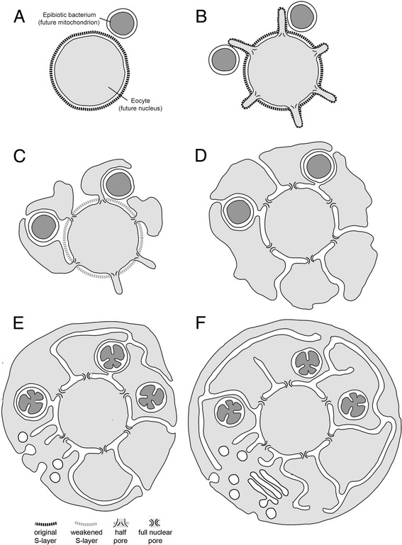
Results: Drawing on diverse aspects of cell biology and phylogenetic data, we invert the traditional interpretation of eukaryotic cell evolution. We propose that an ancestral prokaryotic cell, homologous to the modern-day nucleus, extruded membrane-bound blebs beyond its cell wall. These blebs functioned to facilitate material exchange with ectosymbiotic proto-mitochondria. The cytoplasm was then formed through the expansion of blebs around proto-mitochondria, with continuous spaces between the blebs giving rise to the endoplasmic reticulum, which later evolved into the eukaryotic secretory system. Further bleb-fusion steps yielded a continuous plasma membrane, which served to isolate the endoplasmic reticulum from the environment.

Conclusions: The inside-out theory is consistent with diverse kinds of data and provides an alternative framework by which to explore and understand the dynamic organization of modern eukaryotic cells. It also helps to explain a number of previously enigmatic features of cell biology, including the autonomy of nuclei in syncytia and the subcellular localization of protein N-glycosylation, and makes many predictions, including a novel mechanism of interphase nuclear pore insertion.


Figures

Figure 1
Figure 1
Inside-out model for the evolution of eukaryotic cell organization. Model showing the stepwise evolution of eukaryotic cell organization from (A) an eocyte ancestor with a single bounding membrane and a glycoprotein rich cell wall (S-layer) interacting with epibiotic α-proteobacteria (proto-mitochondria). (B) We envision the eocyte cell forming protrusions, aided by protein-membrane interactions at the protrusion neck. These protrusions facilitated material exchange with proto-mitochondria. (C) Selection for a greater area of contact between the symbionts would have led to bleb enlargement and the eventual loss of the S-layer from the protrusions. (D) Blebs would have then been further stabilized by the development of a symmetric nuclear pore outer ring complex (Figure 2) and through the establishment of LINC complexes that, following the gradual loss of the S-layer, physically connected the original cell body (the nascent nuclear compartment) to the inner bleb membranes. (E) With the expansion of blebs to enclose the proto-mitochondria, a process that would have facilitated the acquisition of bacterial lipid biosynthesis machinery by the host, the site of cell growth would have progressively shifted to the cytoplasm, facilitated by the development of regulated traffic through the nuclear pore. At the same time, the spaces between blebs would have enabled the gradual maturation of proteins secreted into the environment via the perinuclear space through glycosylation and proteolytic cleavage. (F) Finally, bleb fusion would have connected cytoplasmic compartments and driven the formation of an intact plasma membrane, perhaps through a process akin to phagocytosis whereby one bleb enveloped the whole. This simple topological transition would have isolated the endoplasmic reticulum from the outside world, driven the full development of a system of vesicular trafficking, and established strict vertical transmission of mitochondria, leading to a cell with modern eukaryotic cell organization.
Figure 2
Figure 2
Example of epibiotic bacteria associated with archaeal cells. Image of two Candidatus Giganthauma karukerense cells surrounded by ectosymbiotic γ-proteobacteria (reproduced with permission from [84]).
Figure 3
Figure 3
Model for the evolution of nuclear pores and cytoplasmic blebs. (A) Membrane protrusions are formed that extend through holes in the cell wall (S-layer, shown in gray) of the eukaryote ancestor. Protrusions could initially have been coated with an S-layer that was later lost. We propose that protrusions gained structural support at their bases from proteins with seven-blade β-propeller domains (homologs of nucleoporins and COPII coatomers), which stabilize positively curved membranes. Additionally, blebs may have been stabilized by an internal cytoskeleton (red), like that provided by microtubules in modern day flagella, and by components of LINC complexes that connect the cell membrane (and underlying structures) to the S-layer (gray). (B) Lateral spreading of the bleb is aided by the movement of LINC proteins to the inner bleb membrane and by the recruitment of a second, outer ring of nuclear pore proteins to stabilize positive curvature outside of the cell wall.
Figure 4
Figure 4
Model for the evolution of cell division. Cell division is depicted for the ancestral eocyte (A), and at two intermediate stages in the evolution of eukaryotes, before (B) or after (C) bleb fusion. Following the acquisition of blebs, ESCRTIII is used to drive the scission of cytoplasmic bridges connecting cells (likely aided by the archaeal-derived actin cytoskeleton [51]), while LINC complexes and the formation of new nuclear pores restore cell and nuclear organization following division. Mitochondrial segregation is likely aided by host induced Dynamin-mediated scission within the endoplasmic reticulum (not depicted), as observed in modern eukaryotes [91].
Figure 5
Figure 5
The stepwise evolution of eukaryotic vesicle trafficking. From left to right the figure depicts a simple hypothesis for the evolution of the eukaryotic secretion and vesicle trafficking systems. Initially, proteins (black dots) would have been secreted from ribosomes bound to rough endoplasmic reticulum (ER) into the space at the bases of blebs by the Sec translocase and signal recognition particles (SRP) [50]. Secreted proteins could then undergo stepwise processing using machinery adapted from that used to process glycoproteins in the archaeal S-layer (that is, through N-linked glycosylation of asparagine-X-serine or asparagine-X-threonine-containing proteins, and proteolysis [99]). The elaboration of ER tubules and local membrane bending regulated by the Sar1 GTPase, in the presence of generic SNAP Receptors (SNAREs) (blue bars), would have enabled the transient fusion of ER to the outer cell membrane, releasing these glycosylated proteins into the extracellular space. These transient openings would have been closed by Dynamin-mediated fission. Specialized SNARE proteins (differently colored bars) and Dynamin (triple diagonal lines), would then have generated vesicular intermediates to better regulate secretion. The intercalation of additional processing steps and the diversification of these protein families would have yielded compartment-specific paralogs, together with the evolution of regulatory Arf and Rab GTPases, and a Golgi compartment. Finally, membrane bending machinery together with Dynamin, actin, and Rho family GTPases would have been co-opted to drive endocytosis, phagocytosis, and the development of the modern retrograde trafficking pathway.
Figure 6
Figure 6
Comparison of the predicted ordering of cellular innovations, and the corresponding molecular machines, under the inside-out and autogenous outside-in models. Ran, Rab, Sar1, and Rho refer to small GTPase subfamilies. Abbreviations: LINC = Linker of Nucleoskeleton and Cytoskeleton; COPII = Coat protein II; SNAREs = SNAP Receptors.
Figure 7
Figure 7
Predicted mechanism of interphase eukaryotic nuclear pore insertion predicted by the inside-out model. (A) The nuclear envelope is held together through LINC complexes. (B, C) Folds in the inner membrane of the envelope recruit the outer ring of the nuclear pore, composed of proteins with COPII-like domains, to generate a small extranuclear bleb, which is stabilized via the assembly of the complete nuclear pore complex. (D) The nuclear pore complex, together with LINC complexes, generates a tight membrane fold at the bud neck. (E) The nascent bleb is connected to the rest of the cytoplasm by active bleb-bleb fusion, ensuring cytoplasmic continuity. Note that in this model the continuity of the perinuclear space and the endoplasmic reticulum (ER) is a simple consequence of the mechanism of bleb generation. The relative rates at which bleb expansion (A-D) and the fusion of cytoplasmic compartments (E) occur will determine the size of individual cytoplasmic blebs and the extent of cytoplasmic compartmentalization. Thus, if the compartment fusion reaction (D, E) is induced immediately, no enlarged blebs would be seen.

References

    1. Lane N, Martin W. The energetics of genome complexity. Nature. 2010;467:929–934. doi: 10.1038/nature09486. - DOI - PubMed
    1. Cavalier-Smith T. The phagotrophic origin of eukaryotes and phylogenetic classification of protozoa. Int J Syst Evol Microbiol. 2002;52:297–354. doi: 10.1099/00207713-52-2-297. - DOI - PubMed
    1. Koonin EV. The Biological Big Bang model for the major transitions in evolution. Biol Direct. 2007;2:21. doi: 10.1186/1745-6150-2-21. - DOI - PMC - PubMed
    1. Margulis L. Origin of Eukaryotic Cells. New Haven: Yale University Press; 1970.
    1. Margulis L. Symbiosis in Cell Evolution. New York: W. H. Freeman; 1981.