. 2015 Jan 30;116(3):418-24.
doi: 10.1161/CIRCRESAHA.116.304510. Epub 2014 Oct 28.

MicroRNA induced cardiac reprogramming in vivo: evidence for mature cardiac myocytes and improved cardiac function

Affiliations

MicroRNA induced cardiac reprogramming in vivo: evidence for mature cardiac myocytes and improved cardiac function

Tilanthi M Jayawardena et al. Circ Res. .

Abstract

Rationale: A major goal for the treatment of heart tissue damaged by cardiac injury is to develop strategies for restoring healthy heart muscle through the regeneration and repair of damaged myocardium. We recently demonstrated that administration of a specific combination of microRNAs (miR combo) into the infarcted myocardium leads to direct in vivo reprogramming of noncardiac myocytes to cardiac myocytes. However, the biological and functional consequences of such reprogramming are not yet known.

Objective: The aim of this study was to determine whether noncardiac myocytes directly reprogrammed using miRNAs in vivo develop into mature functional cardiac myocytes in situ, and whether reprogramming leads to improvement of cardiac function.

Methods and results: We subjected fibroblast-specific protein 1-Cre mice/tandem dimer Tomato (tdTomato) mice to cardiac injury by permanent ligation of the left anterior descending coronary artery and injected lentiviruses encoding miR combo or a control nontargeting miRNA. miR combo significantly increased the number of reprogramming events in vivo. Five to 6 weeks after injury, morphological and physiological properties of tdTomato(-) and tdTomato(+) cardiac myocyte-like cells were analyzed ex vivo. tdTomato(+) cells expressed cardiac myocyte markers, sarcomeric organization, excitation-contraction coupling, and action potentials characteristic of mature ventricular cardiac myocytes (tdTomato(-) cells). Reprogramming was associated with improvement of cardiac function, as analyzed by serial echocardiography. There was a time delayed and progressive improvement in fractional shortening and other measures of ventricular function, indicating that miR combo promotes functional recovery of damaged myocardium.

Conclusions: The findings from this study further validate the potential use of miRNA-mediated reprogramming as a therapeutic approach to promote cardiac regeneration after myocardial injury.

Keywords: cellular reprogramming; excitation–contraction coupling; guided tissue regeneration; microRNAs; myocardial infarction; myocytes, cardiac.


Conflict of interest statement

DISCLOSURES

None of the authors have any real or apparent conflict(s) of interest to disclose.

Figures
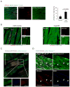
Figure 1
Figure 1. miR combo promotes reprogramming in vivo
FSP1-Cre/tdTomato mice were subjected to either a sham operation or myocardial infarction [MI] and injected with lentivirus containing either miR combo or negmiR. Seven weeks following injury the entire peri-infarct region was visualized by serial sectioning through the heart tissue. Sections were probed for (A) tdTomato and cardiac troponin-T, (B) tdTomato and α-sarcomeric actinin, (C) tdTomato, cardiac troponin-T, and connexin-43, and (D) tdTomato, cardiac troponin-T and DDR2. For all panels, scale bar 100 microns, n=3, p-values indicated.
Figure 2
Figure 2. miR combo reprogrammed cardiac myocytes express mature cardiac markers
Images of purified tdTomato+ cardiac myocytes immunostained for sarcomeric α-actinin, cardiac troponin T, N-cadherin, and Connexin 43 (green), together with tdTomato (red) and DAPI (blue). For each cardiac marker, images shown include overlay of cardiac marker and DAPI (upper), tdTomato and DAPI (middle), and merged image of marker, tdTomato, and DAPI (lower). Scale bar, 100 μm.
Figure 3
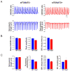
Figure 3. Excitation-contraction coupling in tdTomato+ cardiac myocytes
(A) Representative examples of simultaneous calcium transients and contractility traces obtained from Fura-2 loaded tdTomato and tdTomato+ cardiac myocytes during pacing at 1 Hz with electrical field stimulation. (B) Calcium transient parameters measured using for tdTomato (WT; n = 22) and tdTomato+ (Tom+; n = 12) cardiac myocytes paced at 1 Hz as in A. These include basal Fura-2 ratio (tdTomato = 0.54 ± 0.018; tdTomato+ = 0.53 ± 0.014; p = 0.55) and calcium transient peak amplitude (tdTomato = 0.23 ± 0.021; tdTomato+ = 0.20 ± 0.034; p = 0.47) and decay kinetics (tdTomato = 0.151 ± 0.0088 s; tdTomato+ = 0.133 ± 0.00085 s; p = 0.20). (C) Contractility parameters include basal sarcomeric length (tdTomato = 1.80 ± 0.013; tdTomato+ = 1.84 ± 0.018; p = 0.13), fractional shortening (tdTomato = 5.77 ± 1.08%; tdTomato+ = 4.64 ± 1.08%; p = 0.51), and contraction decay kinetics (tdTomato = 0.063 ± 0.011 s; tdTomato+ = 0.047 ± 0.0053 s; p = 0.31). EC coupling gain (fractional shortening/calcium transient amplitude), reflects the relationship between calcium transient amplitude and contractility (tdTomato = 27.5 ± 5.5; tdTomato+ = 34.4 ± 12.0; p = 0.55). None of these measures were significantly different between tdTomato and tdTomato+ cardiac myocytes.
Figure 4
Figure 4. Electrophysiological properties of tdTomato+ cardiac myocytes
(A) Representative recordings of action potentials evoked in tdTomato and tdTomato+ cardiac myocytes. (B) Table of action potential parameters measured for tdTomato (n = 11) and tdTomato+ (n = 7) cardiac myocytes including RMP, APA, OSP, and action potential duration (APD) at 30 and 70% repolarization. P values, listed for each measure, were < 0.05 for RMP, OSP, and APA. (C) Examples of I-V relationship in a tdTomato and a tdTomato+ cardiomyocyte. (D) Average I-V curves in tdTomato (n = 6) and tdTomato+ (n = 3) cardiac myocytes generated from steady-state current at the end of the voltage step. There was no statistically significant difference in current amplitude at any voltage step potential.
Figure 5
Figure 5. miR combo delivery improves cardiac function following myocardial infarction
Cardiac function was assessed in mice injected with negmiR (n = 9) or miR combo (n = 6) by echocardiographic analysis before and at 2 weeks and 1, 2, and 3 months post-infarct. (A) Fractional shortening, Vcfc, LV mass, and LVEDS of miR combo vs negmiR mice. * indicates p < 0.05 compared to negmiR group as determined by ANOVA. (B) Distribution of fractional shortening results of individual mice among miR combo and negmiR-injected groups at 2 months (negmiR = 18.3 ± 2.3%; miR combo = 25.7 ± 2.2%; p = 0.045) and 3 months (negmiR = 19.8 ± 1.9%; miR combo = 30.8 ± 4.1%; p = 0.019) post-MI. Mean is indicated by black horizontal bar and SEM is indicated by blue horizontal bars. (C) Serial sections from miR combo and negmiR mice were stained with Masson’s trichrome one month following injury. Fibrosis area is representated as a percentage of the left ventricle. N = 6 miR combo, n = 5 negmiR.

Comment in

  • Small RNAs make big impact in cardiac repair.
    Krane M, Deutsch MA, Doppler S, Lange R, Wu SM. Krane M, et al. Circ Res. 2015 Jan 30;116(3):393-5. doi: 10.1161/CIRCRESAHA.114.305676. Circ Res. 2015. PMID: 25634967 Free PMC article. No abstract available.

References

    1. Heidenreich PA, Trogdon JG, Khavjou OA, Butler J, Dracup K, Ezekowitz MD, Finkelstein EA, Hong Y, Johnston SC, Khera A, Lloyd-Jones DM, Nelson SA, Nichol G, Orenstein D, Wilson PW, Woo YJ. Forecasting the future of cardiovascular disease in the united states: A policy statement from the american heart association. Circulation. 2011;123:933–944. - PubMed
    1. Ieda M, Fu JD, Delgado-Olguin P, Vedantham V, Hayashi Y, Bruneau BG, Srivastava D. Direct reprogramming of fibroblasts into functional cardiac myocytes by defined factors. Cell. 2010;142:375–386. - PMC - PubMed
    1. Qian L, Huang Y, Spencer CI, Foley A, Vedantham V, Liu L, Conway SJ, Fu JD, Srivastava D. In vivo reprogramming of murine cardiac fibroblasts into induced cardiac myocytes. Nature. 2012;485:593–598. - PMC - PubMed
    1. Song K, Nam YJ, Luo X, Qi X, Tan W, Huang GN, Acharya A, Smith CL, Tallquist MD, Neilson EG, Hill JA, Bassel-Duby R, Olson EN. Heart repair by reprogramming non-myocytes with cardiac transcription factors. Nature. 2012;485:599–604. - PMC - PubMed
    1. Addis RC, Epstein JA. Induced regeneration--the progress and promise of direct reprogramming for heart repair. Nat Med. 2013;19:829–836. - PMC - PubMed

Publication types