. 2015 Jan 15;465(2):271-9.
doi: 10.1042/BJ20141165.

The clinically approved drugs dasatinib and bosutinib induce anti-inflammatory macrophages by inhibiting the salt-inducible kinases

Affiliations

The clinically approved drugs dasatinib and bosutinib induce anti-inflammatory macrophages by inhibiting the salt-inducible kinases

James Ozanne et al. Biochem J. .

Abstract

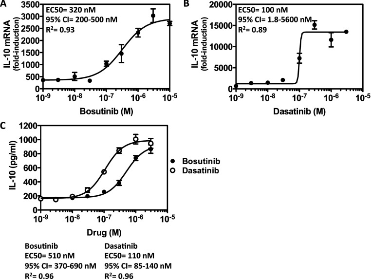
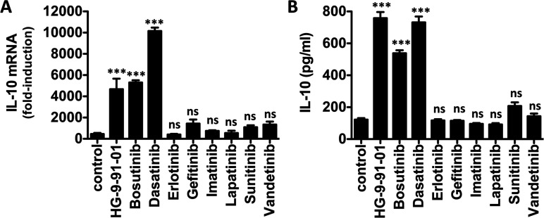
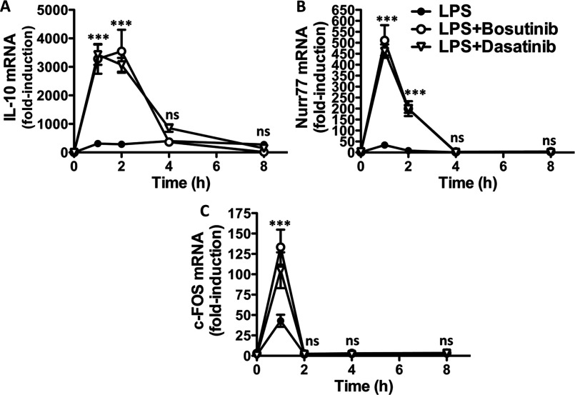
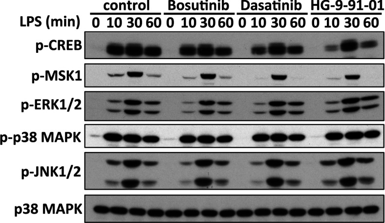
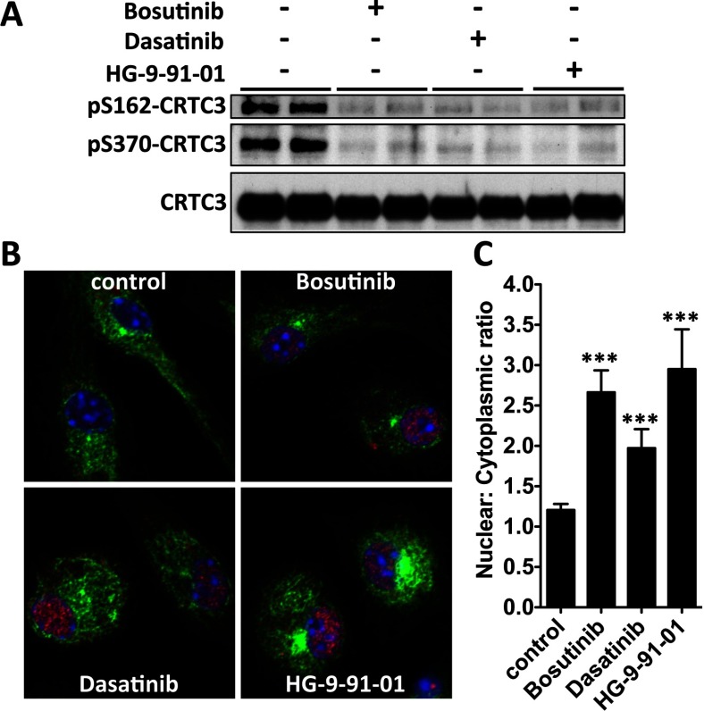
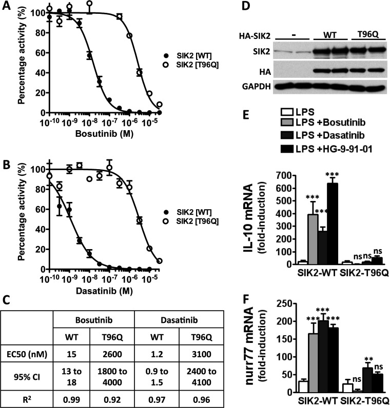
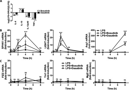
Macrophages switch to an anti-inflammatory, 'regulatory'-like phenotype characterized by the production of high levels of interleukin (IL)-10 and low levels of pro-inflammatory cytokines to promote the resolution of inflammation. A potential therapeutic strategy for the treatment of chronic inflammatory diseases would be to administer drugs that could induce the formation of 'regulatory'-like macrophages at sites of inflammation. In the present study, we demonstrate that the clinically approved cancer drugs bosutinib and dasatinib induce several hallmark features of 'regulatory'-like macrophages. Treatment of macrophages with bosutinib or dasatinib elevates the production of IL-10 while suppressing the production of IL-6, IL-12p40 and tumour necrosis factor α (TNFα) in response to Toll-like receptor (TLR) stimulation. Moreover, macrophages treated with bosutinib or dasatinib express higher levels of markers of 'regulatory'-like macrophages including LIGHT, SPHK1 and arginase 1. Bosutinib and dasatinib were originally developed as inhibitors of the protein tyrosine kinases Bcr-Abl and Src but we show that, surprisingly, the effects of bosutinib and dasatinib on macrophage polarization are the result of the inhibition of the salt-inducible kinases. Consistent with the present finding, bosutinib and dasatinib induce the dephosphorylation of CREB-regulated transcription co-activator 3 (CRTC3) and its nuclear translocation where it induces a cAMP-response-element-binding protein (CREB)-dependent gene transcription programme including that of IL-10. Importantly, these effects of bosutinib and dasatinib on IL-10 gene expression are lost in macrophages expressing a drug-resistant mutant of salt-inducible kinase 2 (SIK2). In conclusion, our study identifies the salt-inducible kinases as major targets of bosutinib and dasatinib that mediate the effects of these drugs on the innate immune system and provides novel mechanistic insights into the anti-inflammatory properties of these drugs.


Figures

Figure 1
Figure 1. Bosutinib and dasatinib elevate IL-10 mRNA production and secretion in a dose-dependent manner
Bone-marrow-derived macrophages were treated with the indicated concentrations of (A) bosutinib and (B) dasatinib for 1 h and then stimulated with LPS for 1 h. IL-10 mRNA levels were measured by qPCR (n=4, mean±S.E.M.). (C) Experiment was performed as above except culture supernatants were harvested after stimulation with LPS for 2 h. The concentration of IL-10 secreted into the culture medium was measured using the BIOPLEX system (n=4, mean±S.E.M.).
Figure 2
Figure 2. Bosutinib and dasatinib but not other protein tyrosine kinase inhibitors elevate IL-10 production in macrophages
Bone-marrow-derived macrophages were treated with vehicle control or the indicated compounds for 1 h and then stimulated with LPS for (A) 1 h to induce mRNA expression or (B) 2 h for cytokine secretion. IL-10 mRNA levels were measured by qPCR, whereas the concentration of IL-10 secreted into the culture medium was measured using the BIOPLEX system (n=4, mean±S.E.M.). All compounds were used at 10 μM except HG-9-91-01, bosutinib and dasatinib which were used at 0.5, 3 and 0.3 μM respectively.
Figure 3
Figure 3. Bosutinib and dasatinib induce a gene expression programme directed by the transcription factor CREB
Bone-marrow-derived macrophages were treated with vehicle control, 3 μM bosutinib or 0.3 μM dasatinib for 1 h and then stimulated with LPS for the indicated times. Expression of different CREB-dependent genes, (A) IL-10, (B) nurr77 and (C) c-FOS, were measured by qPCR. mRNA levels were normalized to 1 in unstimulated cells (mean±S.E.M., n=4).
Figure 4
Figure 4. Bosutinib and dasatinib do not affect the TLR-stimulated phosphorylation of CREB
Bone-marrow-derived macrophages were treated with vehicle control, 3 μM bosutinib, 0.3 μM dasatinib or 0.5 μM HG-9-91-01 for 1 h and then stimulated with LPS for 0, 10, 30, or 60 min. Cell lysates were immunoblotted using the indicated antibodies.
Figure 5
Figure 5. Bosutinib and dasatinib induce the dephosphorylation of CRTC3 and its nuclear translocation
(A) RAW264.7 macrophages were treated with bosutinib, dasatinib, HG-9-91-01 or vehicle control for 1 h. CRTC3 was immunoprecipitated and phosphorylation of Ser162 and Ser370 was monitored by immunoblotting. (B) Bone-marrow-derived macrophages were plated on glass coverslips, and treated with bosutinib, dasatinib, HG-9-91-01 or vehicle control for 1 h. Samples were stained for CRTC3 (Alexa Fluor® 594), tubulin (Alexa Fluor® 488) and DNA (DAPI) content and imaged under a confocal microscope. (C) Quantification of CRTC3 nuclear translocation as a function of its nuclear to cytoplasmic ratio. (mean±S.E.M., n=10).
Figure 6
Figure 6. Bosutinib and dasatinib fail to induce IL-10 production in macrophages expressing a drug-resistant mutant of SIK2
The SIK2[T96Q] mutant is insensitive to (A) bosutinib and (B) dasatinib in vitro. Kinase assays were performed using purified SIK2 wild-type and SIK2[T96Q] in the presence of increasing amounts of inhibitors as described in the Materials and methods section (mean±S.D., n=2). (C) Statistical analysis of IC50 curves generated in (A) and (B). (D) RAW264.7 macrophages engineered to express HA-SIK2-WT and HA-SIK2[T96Q] were treated with doxycycline for 16 h, lysed and immunoblotted with the indicated antibodies. Cells transduced with a retrovirus containing no insert was used as negative control (−). (E and F) RAW264.7 macrophages expressing HA-SIK2-WT and HA-SIK2[T96Q] were treated with bosutinib, dasatinib, HG-9-91-01 or vehicle control for 1 h and then stimulated with LPS for 1 h. mRNA expression of IL-10 (E) and nurr77 (F) were measured by qPCR (mean±S.E.M., n=4). WT, wild-type.
Figure 7
Figure 7. Bosutinib and dasatinib induce key features of ‘regulatory’ macrophages
(A) Effect of bosutinib and dasatinib on cytokine secretion. Bone-marrow-derived macrophages were treated with vehicle control, 3 μM bosutinib or 0.3 μM dasatinib for 1 h and then stimulated with LPS for 8 h. The concentration of the different cytokines in the culture supernatant was measured using the BIOPLEX system. The data are depicted as the fold-change in cytokine secretion in the presence of the drug (n=4, mean±S.E.M.). Statistical significance was determined by comparing each dataset to 0 using a one-sample Student's t test. (B) Effect of bosutinib and dasatinib on the expression of markers of ‘regulatory’ macrophages. Bone-marrow-derived macrophages were treated with vehicle control, 3 μM bosutinib or 0.3 μM dasatinib for 1 h and then stimulated with LPS for the indicated times. Expression of LIGHT, SPHK1 and arginase 1 were measured by qPCR. mRNA levels were normalized to 1 in unstimulated cells (mean±S.E.M., n=4). (C) Effect of bosutinib and dasatinib on the expression of markers of alternatively activated macrophages. Experiment was performed as described in (B) but measuring the mRNA levels of FIZZ, Ym1 and Mgl2 (mean±S.E.M., n=4).

References

    1. Serhan C. N., Savill J. Resolution of inflammation: the beginning programs the end. Nat. Immunol. 2005;6:1191–1197. doi: 10.1038/ni1276. - DOI - PubMed
    1. Sica A., Mantovani A. Macrophage plasticity and polarization: in vivo veritas. J. Clin. Invest. 2012;122:787–795. doi: 10.1172/JCI59643. - DOI - PMC - PubMed
    1. Martinez F. O., Gordon S. The M1 and M2 paradigm of macrophage activation: time for reassessment. F1000Prime Rep. 2014;6:13. doi: 10.12703/P6-13. - DOI - PMC - PubMed
    1. Mosser D. M., Edwards J. P. Exploring the full spectrum of macrophage activation. Nat. Rev. Immunol. 2008;8:958–969. doi: 10.1038/nri2448. - DOI - PMC - PubMed
    1. Edwards J. P., Zhang X., Frauwirth K. A., Mosser D. M. Biochemical and functional characterization of three activated macrophage populations. J. Leuk. Biol. 2006;80:1298–1307. doi: 10.1189/jlb.0406249. - DOI - PMC - PubMed

Publication types

MeSH terms

Substances