

Syrian hamsters (Mesocricetus auratus) oronasally inoculated with a Nipah virus isolate from Bangladesh or Malaysia develop similar respiratory tract lesions
- PMID: 25352203
- PMCID: PMC6309986
- DOI: 10.1177/0300985814556189
Syrian hamsters (Mesocricetus auratus) oronasally inoculated with a Nipah virus isolate from Bangladesh or Malaysia develop similar respiratory tract lesions
Abstract
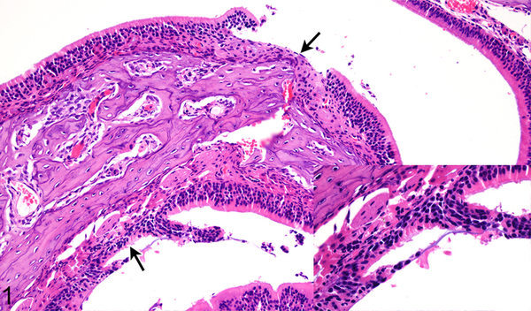
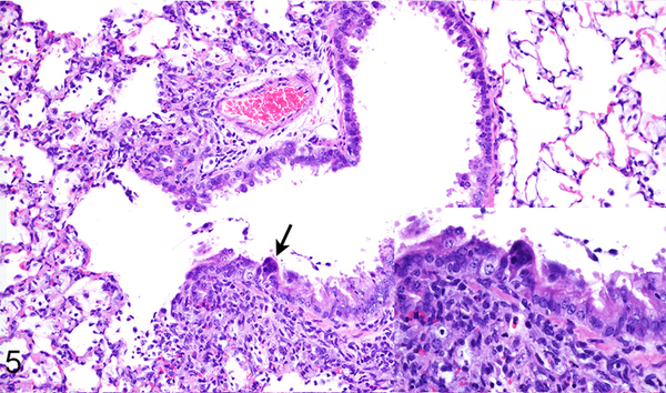
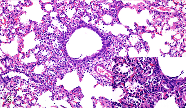
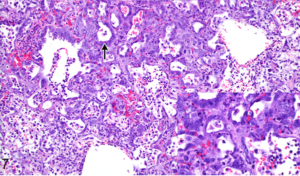
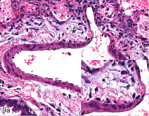
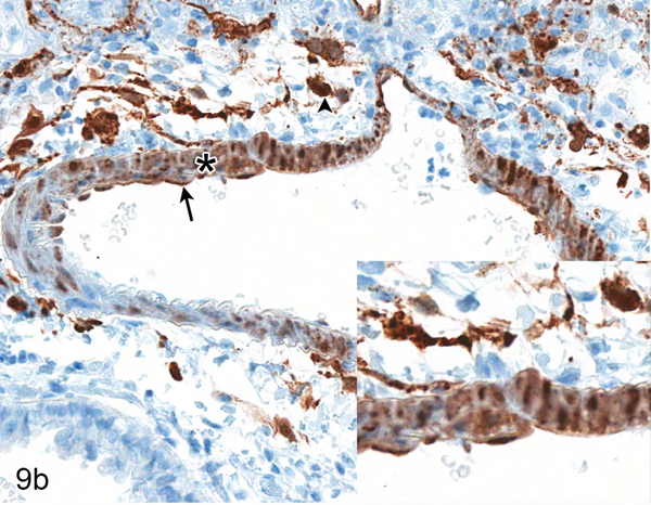
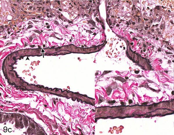
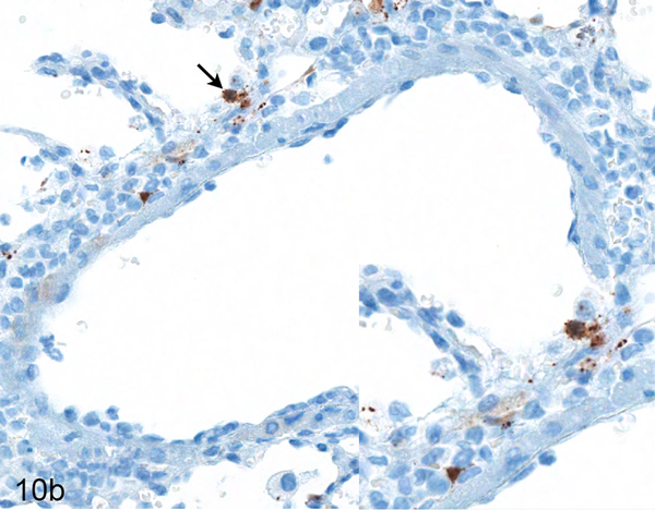
Nipah virus is a paramyxovirus in the genus Henipavirus, which has caused outbreaks in humans in Malaysia, India, Singapore, and Bangladesh. Whereas the human cases in Malaysia were characterized mainly by neurological symptoms and a case fatality rate of ∼40%, cases in Bangladesh also exhibited respiratory disease and had a case fatality rate of ∼70%. Here, we compared the histopathologic changes in the respiratory tract of Syrian hamsters, a well-established small animal disease model for Nipah virus, inoculated oronasally with Nipah virus isolates from human cases in Malaysia and Bangladesh. The Nipah virus isolate from Bangladesh caused slightly more severe rhinitis and bronchointerstitial pneumonia 2 days after inoculation in Syrian hamsters. By day 4, differences in lesion severity could no longer be detected. Immunohistochemistry demonstrated Nipah virus antigen in the nasal cavity and pulmonary lesions; the amount of Nipah virus antigen present correlated with lesion severity. Immunohistochemistry indicated that both Nipah virus isolates exhibited endotheliotropism in small- and medium-caliber arteries and arterioles, but not in veins, in the lung. This correlated with the location of ephrin B2, the main receptor for Nipah virus, in the vasculature. In conclusion, Nipah virus isolates from outbreaks in Malaysia and Bangladesh caused a similar type and severity of respiratory tract lesions in Syrian hamsters, suggesting that the differences in human disease reported in the outbreaks in Malaysia and Bangladesh are unlikely to have been caused by intrinsic differences in these 2 virus isolates.
Keywords: Nipah virus; Syrian hamster; artery; histopathology; pathogenicity; respiratory system; tropism.
© The Author(s) 2014.
Figures
Similar articles
-
Comparison of the pathogenicity of Nipah virus isolates from Bangladesh and Malaysia in the Syrian hamster.PLoS Negl Trop Dis. 2013;7(1):e2024. doi: 10.1371/journal.pntd.0002024. Epub 2013 Jan 17. PLoS Negl Trop Dis. 2013. PMID: 23342177 Free PMC article.
-
Identifying Early Target Cells of Nipah Virus Infection in Syrian Hamsters.PLoS Negl Trop Dis. 2016 Nov 3;10(11):e0005120. doi: 10.1371/journal.pntd.0005120. eCollection 2016 Nov. PLoS Negl Trop Dis. 2016. PMID: 27812087 Free PMC article.
-
Intracranial inoculation rapidly induces Nipah virus encephalitis in Syrian hamsters.PLoS Negl Trop Dis. 2024 Oct 28;18(10):e0012635. doi: 10.1371/journal.pntd.0012635. eCollection 2024 Oct. PLoS Negl Trop Dis. 2024. PMID: 39466844 Free PMC article.
-
The emergence of Nipah virus, a highly pathogenic paramyxovirus.J Clin Virol. 2008 Dec;43(4):396-400. doi: 10.1016/j.jcv.2008.08.007. Epub 2008 Oct 2. J Clin Virol. 2008. PMID: 18835214 Review.
-
Nipah Virus Infection.J Clin Microbiol. 2018 May 25;56(6):e01875-17. doi: 10.1128/JCM.01875-17. Print 2018 Jun. J Clin Microbiol. 2018. PMID: 29643201 Free PMC article. Review.
Cited by
-
Pathogenicity and virulence of henipaviruses.Virulence. 2023 Dec;14(1):2273684. doi: 10.1080/21505594.2023.2273684. Epub 2023 Nov 10. Virulence. 2023. PMID: 37948320 Free PMC article. Review.
-
Henipavirus Immune Evasion and Pathogenesis Mechanisms: Lessons Learnt from Natural Infection and Animal Models.Viruses. 2022 Apr 29;14(5):936. doi: 10.3390/v14050936. Viruses. 2022. PMID: 35632678 Free PMC article. Review.
-
Contribution of Human Lung Parenchyma and Leukocyte Influx to Oxidative Stress and Immune System-Mediated Pathology following Nipah Virus Infection.J Virol. 2017 Jul 12;91(15):e00275-17. doi: 10.1128/JVI.00275-17. Print 2017 Aug 1. J Virol. 2017. PMID: 28539439 Free PMC article.
-
Pathogenic Differences between Nipah Virus Bangladesh and Malaysia Strains in Primates: Implications for Antibody Therapy.Sci Rep. 2016 Aug 3;6:30916. doi: 10.1038/srep30916. Sci Rep. 2016. PMID: 27484128 Free PMC article.
-
Henipavirus Encephalitis: Recent Developments and Advances.Brain Pathol. 2015 Sep;25(5):605-13. doi: 10.1111/bpa.12278. Brain Pathol. 2015. PMID: 26276024 Free PMC article. Review.
References
-
- Centers for Disease Control and Prevention. Outbreak of Hendra-like virus - Malaysia and Singapore, 1998–1999. MMWR Morb Mortal Wkly Rep. 1999;48:265–269. - PubMed
-
- Chew MH, Arguin PM, Shay DK, et al. Risk factors for Nipah virus infection among abattoir workers in Singapore. J Infect Dis. 2000;181:1760–1763. - PubMed
-
- Chua KB, Bellini WJ, Rota PA, et al. Nipah virus: a recently emergent deadly paramyxovirus. Science. 2000;288:1432–1435. - PubMed
Publication types
MeSH terms
Grants and funding
LinkOut - more resources
Full Text Sources
Other Literature Sources
