Review
. 2014 Dec;10(4):529-64.
doi: 10.1007/s11302-014-9427-2. Epub 2014 Oct 29.

Purinergic signalling and immune cells

Affiliations
Review

Purinergic signalling and immune cells

Geoffrey Burnstock et al. Purinergic Signal. 2014 Dec.

Abstract

This review article provides a historical perspective on the role of purinergic signalling in the regulation of various subsets of immune cells from early discoveries to current understanding. It is now recognised that adenosine 5'-triphosphate (ATP) and other nucleotides are released from cells following stress or injury. They can act on virtually all subsets of immune cells through a spectrum of P2X ligand-gated ion channels and G protein-coupled P2Y receptors. Furthermore, ATP is rapidly degraded into adenosine by ectonucleotidases such as CD39 and CD73, and adenosine exerts additional regulatory effects through its own receptors. The resulting effect ranges from stimulation to tolerance depending on the amount and time courses of nucleotides released, and the balance between ATP and adenosine. This review identifies the various receptors involved in the different subsets of immune cells and their effects on the function of these cells.


Figures
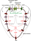
Fig. 1
Fig. 1
Proposed model of neutrophil chemotaxis. As previously reported, stimulation of chemoattractant receptors induces local release of ATP through PANX1 channels at the site that first encounters the chemoattractant. Autocrine feedback via P2Y2 receptors amplifies the chemotactic signal and triggers cell polarization, whereby cells assume an elongated shape, and PANX1, CD39 (NTPDase1) and A3 adenosine receptors accumulate at the leading edge. In the current study, we found that A2A receptors are translocated from the leading edge toward the back of polarized neutrophils and that inhibitory signaling via A2A receptor-dependent cAMP accumulation inhibits excitatory chemotactic signalling by blocking FPR-dependent ERK and p38 MAPK activation globally with the exception of the leading edge. ALP alkaline phosphatase, ADO adenosine, PIP3 phosphatidylinositol (3,4,5)-triphosphate. (Reproduced from [55], with permission from the American Society for Biochemistry and Molecular Biology)
Fig. 2
Fig. 2
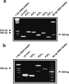
P2 receptors expressed by human eosinophils. a P2Y receptors. b P2X receptors. (Reproduced from [110], with permission from Elsevier)
Fig. 3
Fig. 3
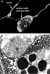
a Close apposition between rat mast cell protease 1 immunoreactive and calcitonin gene-related peptide immunoreactive nerve fibres observed by confocal microscopy. b Ultrathin section of rabbit middle cerebral artery showing granular cells (G) separated by a distance of less than 200 nm. V varicosities; arrowheads basement membranes. Magnification, ×29374. (a Reproduced from [176] and b from [173], with permission from Elsevier)
Fig. 4
Fig. 4
Hypothetical sequence of events leading to P2X7 receptor and pannexin 1 (panx-1)-mediated inflammasome activation. Pathogen-associated molecular patterns (PAMPs) bind to Toll-like receptors (TLRs) and drive interleukin (IL)-1β gene expression and accumulation of the pro-cytokine. Extracellular ATP binds to the P2X7 receptor and triggers K+ efflux and panx-1 activation. The functional significance of K+ efflux is unknown, although it might facilitate or even precipitate inflammasome activation. Likewise, the mechanism of panx-1 activation by the P2X7 receptor is unknown. Panx-1 in turn activates the inflammasome. Data suggest that the ion-carrying activity of panx-1 is unnecessary for inflammasome activation. The activated inflammasome then cleaves pro-IL-1β. Thus, stimulation of the inflammasome by extracellular ATP can be split into two steps: (a) recruitment and activation of panx-1 by the P2X7 receptor and (b) activation of the inflammasome by panx-1. Colour coding: white PAMP, red TLR, green NALP3 inflammasome, orange protein–protein interaction domains, further subdivided into orange square, ASC apoptosis-associated speck-like protein containing a caspase-recruitment domain and orange octagon, pyrin domain; yellow FIIND domain, light blue caspase domain (Casp-1), dark blue biologically active IL-1β and IL-1β propiece, violet P2X7 receptor, light green panx-1. (Reproduced from [210], with permission from Elsevier)
Fig. 5
Fig. 5
Independence of P2Y12 receptor-mediated migration and P2Y6 receptor-mediated phagocytosis in microglia. a Release/leakage of adenine nucleotides/nucleosides and uridine nucleotides from injured neurons. When neurons or cells are injured or dead, high concentrations of ATP (∼mM) and UTP at a concentration of less than 10 % are leaked. Compared with ATP/ADP/adenosine, UTP/UDP should be transient and localized signals. b Changes in P2Y12 and P2Y6 receptors in microglia according to their activation stages. Insert shows pharmacological characterization of P2Y6 receptor. UDP is a selective agonist to the P2Y6 receptor, and thus, it does not stimulate P2Y12, P2X4, A1, or A2A receptors. Similarly, the P2Y6 receptor is a very selective receptor for UDP, and therefore, is not activated by ATP, ADP, or adenosine (Ado). Resting microglia express no or only faint P2Y6 receptors; whereas, they express P2Y12 receptors adequately. When microglia are activated, they increase P2Y6 receptors; whereas, they decrease P2Y12 receptors. Only when activated microglia meet UDP at the injured sites do they sense UDP as an eat-me signal. c Microglial migration and phagocytosis are controlled by distinct P2 receptors. When microglia sense ATP/ADP by P2Y12 receptors, they extrude their processes, followed by migration toward the injured sites. These microglial motilities are not affected by UDP/P2Y6 receptors. When activated, microglia upregulate P2Y6 receptors, and if they sense the eat-me signal UDP, they start to phagocytose the dead cells or debris. The phagocytic responses are not affected by the activation of P2Y12 receptors nor by other P2 or P1 receptors. (Reproduced from [344], with permission from Springer)
Fig. 6
Fig. 6
The CD39/CD73 pathway modulates regulatory T cell (Treg) activity. The activation of T cell receptor (TCR), expressed on Tregs, induces CD39 activity. This increment of ATP-metabolizing activity is critical for the immunosuppressive activity of Tregs because it facilitates the pericellular generation of adenosine, a substantial component of the immunosuppressive and anti-inflammatory functions of Tregs. The inhibitory action of Treg-derived adenosine can be ascribed to the activation of A2A receptors expressed on T effector cells, which undergo reduced immune activity. In addition, adenosine generation triggers a self-reinforcing loop of Treg functions because the stimulation of A2A receptors expressed on these cells elicits their expansion and increases their immunoregulatory activity. (Reproduced from [426], with permission from Elsevier)
Fig. 7
Fig. 7
Purinergic signalling in T cell activation. Antigen recognition by T cells involves the formation of an immune synapse between a T cell and an antigen-presenting cell (APC). The immune synapse contains a large number of signalling molecules that are required for T cell activation, including T cell receptors (TCRs), MHC molecules, co-stimulatory receptors and the purinergic signalling receptors P2X1, P2X4 and P2X7. In response to TCR and CD28 stimulation, pannexin 1, P2X1 receptors and P2X4 receptors translocate to the immune synapse. ATP released through pannexin 1 promotes autocrine signalling via the P2X receptors. Confinement of ATP in the immune synapse results in a powerful autocrine feedback mechanism that facilitates the signal amplification required for antigen recognition. P2 receptors expressed and ATP released by APCs may also have important roles in regulating the antigen recognition process. NFAT nuclear factor of activated T cells. (Reproduced from [474], with permission from Springer)

References

    1. Idzko M, Hammad H, van Nimwegen M, Kool M, Willart MA, Muskens F, Hoogsteden HC, Luttmann W, Ferrari D, Di Virgilio F, Virchow JC, Jr, Lambrecht BN. Extracellular ATP triggers and maintains asthmatic airway inflammation by activating dendritic cells. Nat Med. 2007;13:913–919. - PubMed
    1. Di Virgilio F, Boeynaems JM, Robson SC. Extracellular nucleotides as negative modulators of immunity. Curr Opin Pharmacol. 2009;9:507–513. - PMC - PubMed
    1. Elliott MR, Chekeni FB, Trampont PC, Lazarowski ER, Kadl A, Walk SF, Park D, Woodson RI, Ostankovich M, Sharma P, Lysiak JJ, Harden TK, Leitinger N, Ravichandran KS. Nucleotides released by apoptotic cells act as a find-me signal to promote phagocytic clearance. Nature. 2009;461:282–286. - PMC - PubMed
    1. Homolya L, Steinberg TH, Boucher RC. Cell to cell communication in response to mechanical stress via bilateral release of ATP and UTP in polarized epithelia. J Cell Biol. 2000;150:1349–1360. - PMC - PubMed
    1. McNamara N, Khong A, McKemy D, Caterina M, Boyer J, Julius D, Basbaum C. ATP transduces signals from ASGM1, a glycolipid that functions as a bacterial receptor. Proc Natl Acad Sci U S A. 2001;98:9086–9091. - PMC - PubMed