Review
. 2014 Oct 29:9:43.
doi: 10.1186/1750-1326-9-43.

Alpha-synuclein and tau: teammates in neurodegeneration?

Affiliations
Review

Alpha-synuclein and tau: teammates in neurodegeneration?

Simon Moussaud et al. Mol Neurodegener. .

Abstract

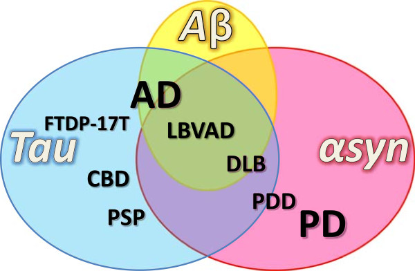
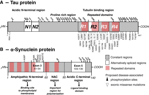
The accumulation of α-synuclein aggregates is the hallmark of Parkinson's disease, and more generally of synucleinopathies. The accumulation of tau aggregates however is classically found in the brains of patients with dementia, and this type of neuropathological feature specifically defines the tauopathies. Nevertheless, in numerous cases α-synuclein positive inclusions are also described in tauopathies and vice versa, suggesting a co-existence or crosstalk of these proteinopathies. Interestingly, α-synuclein and tau share striking common characteristics suggesting that they may work in concord. Tau and α-synuclein are both partially unfolded proteins that can form toxic oligomers and abnormal intracellular aggregates under pathological conditions. Furthermore, mutations in either are responsible for severe dominant familial neurodegeneration. Moreover, tau and α-synuclein appear to promote the fibrillization and solubility of each other in vitro and in vivo. This suggests that interactions between tau and α-synuclein form a deleterious feed-forward loop essential for the development and spreading of neurodegeneration. Here, we review the recent literature with respect to elucidating the possible links between α-synuclein and tau.


Figures

Figure 1
Figure 1
Overlap of proteinopathies. In numerous neurodegenerative disorders, amyloid deposits composed of α-synuclein protein (red circle), tau protein (blue circle) and Aβ peptide (yellow circle) are found. Histopathological classification of neurodegenerative diseases is based on the nature and localization of these deposits in the nervous system. The pathologies are not hermetically isolated categories but form a continuum and concomitance of αsyn and tau pathology is not rare. αSyn pathology (or synucleinopathy) is not restricted to PD but is a feature of several dementing disorders such as PDD, DLB, and frequently occurs in AD where it contributes to secondary symptoms. By contrast tauopathy is repeatedly observed in numerous disorders primarily classified as synucleinopathies and may contribute to clinical heterogeneity.
Figure 2
Figure 2
Schematic representation of tau and α-synuclein proteins. A- Alternative splicing of the N1, N1 + N2 and R2 regions (white) yields in 6 different tau isoforms referred to as 0N3R (=tau23 or tau-352), 0N4R (=tau24 or tau-383), 1N3R (=tau37 or tau-381), 1N4R (=tau46 or tau-412), 2N3R (=tau39 or tau-410) and 2N4R (=tau40 or tau-441). Tau has an acidic N-terminus and a tubulin binding region where the vast majority of the exonic (▽) and intronic (not depicted here) disease-associated mutations are found. B- αSyn is a 14.5 kDa protein divided into 3 major regions; the amphipathic N-terminus, the hydrophobic Non-Amyloid Component (NAC) domain, and the acidic C-terminus. Pathogenic missense mutations described to date (▽) are located in the N-terminal region, whereas most disease-related phosphorylation sites (▲) are localized to the C-terminal region of the protein.
Figure 3
Figure 3
Tau and α-synuclein pathologies. A to D- Histological sections from the amygdala of DLB patients immunostained with an antibody against phosphorylated tau (PHF-1, Abcam, #ab66275) (A and B) or an antibody against phosphorylated αsyn (pSyn#64, Wako, #015-25191) (C and D). Abnormal proteinaceous inclusions of phosphorylated tau protein, called neurofibrillary tangles (A and B, formula image 
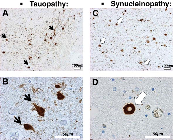
), and of αsyn protein, called Lewy bodies and neurites (C and D, formula image ) are often found in neurons of the amygdala in DLB patients.
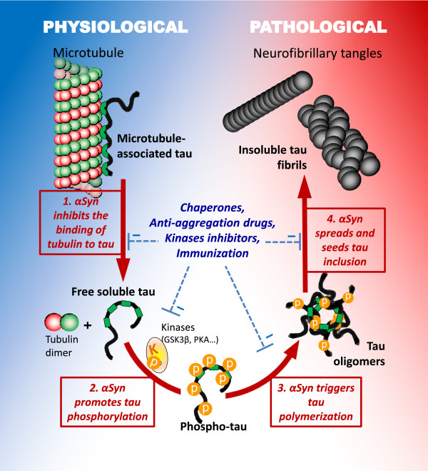
Figure 4
Figure 4
Putative pathways of deleterious tau and α-synuclein interactions. Interaction of tau and αsyn may promote pathogenesis via distinct mechanisms. 1. αSyn may block the normal interaction between tau and tubulin by directly binding to tau and tubulin and thereby interfering with tau physiological function. 2. αSyn could recruit kinases and promote the hyperphosphorylation of tau. 3. αSyn may also catalyze tau polymerization and trigger the formation of tau/αsyn co-oligomers. 4. Finally, αsyn oligomers or fibrils may seed tau fibrillization and thereby initiate and propagate tauopathy.

References

    1. Weintraub D, Comella CL, Horn S. Parkinson's disease–part 1: pathophysiology, symptoms, burden, diagnosis, and assessment. Am J Manag Care. 2008;14:S40–S48. - PubMed
    1. Weintraub D, Comella CL, Horn S. Parkinson's disease–part 2: treatment of motor symptoms. Am J Manag Care. 2008;14:S49–S58. - PubMed
    1. Braak H, Rub U, Gai WP, Del Tredici K. Idiopathic Parkinson's disease: possible routes by which vulnerable neuronal types may be subject to neuroinvasion by an unknown pathogen. J Neural Transm. 2003;110:517–536. doi: 10.1007/s00702-002-0808-2. - DOI - PubMed
    1. Jones DR, Moussaud S, McLean P. Targeting heat shock proteins to modulate alpha-synuclein toxicity. Ther Adv Neurol Disord. 2014;7:33–51. doi: 10.1177/1756285613493469. - DOI - PMC - PubMed
    1. Spillantini MG, Schmidt ML, Lee VM, Trojanowski JQ, Jakes R, Goedert M. Alpha-synuclein in Lewy bodies. Nature. 1997;388:839–840. doi: 10.1038/42166. - DOI - PubMed

Publication types

MeSH terms