. 2014 Nov 10;42(20):12789-805.
doi: 10.1093/nar/gku952. Epub 2014 Oct 28.

A cytoplasmic RNA virus generates functional viral small RNAs and regulates viral IRES activity in mammalian cells

Affiliations

A cytoplasmic RNA virus generates functional viral small RNAs and regulates viral IRES activity in mammalian cells

Kuo-Feng Weng et al. Nucleic Acids Res. .

Abstract

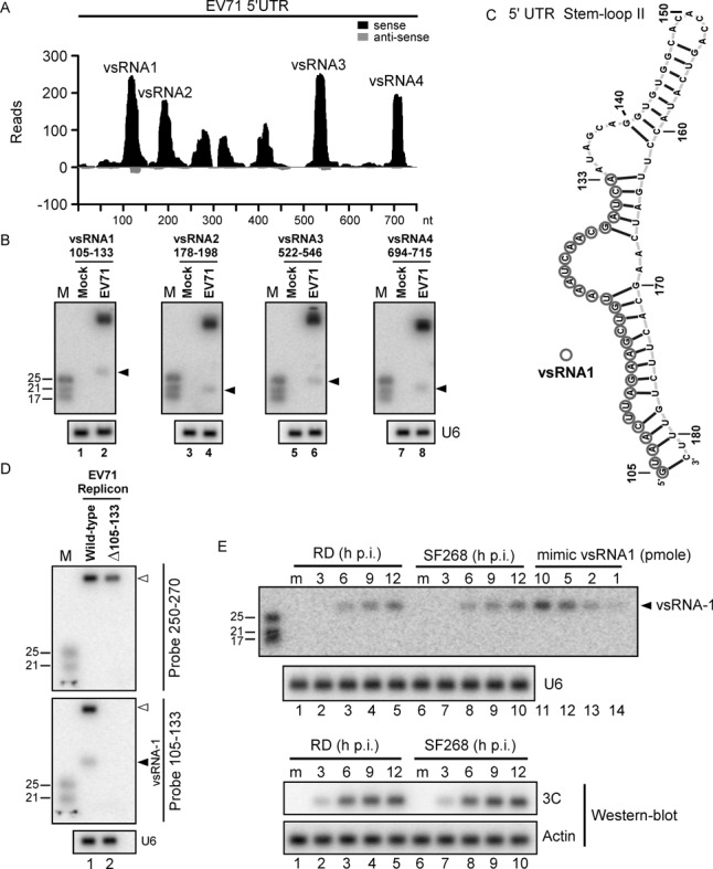
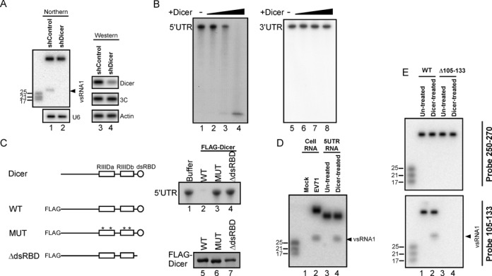
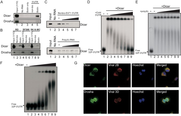
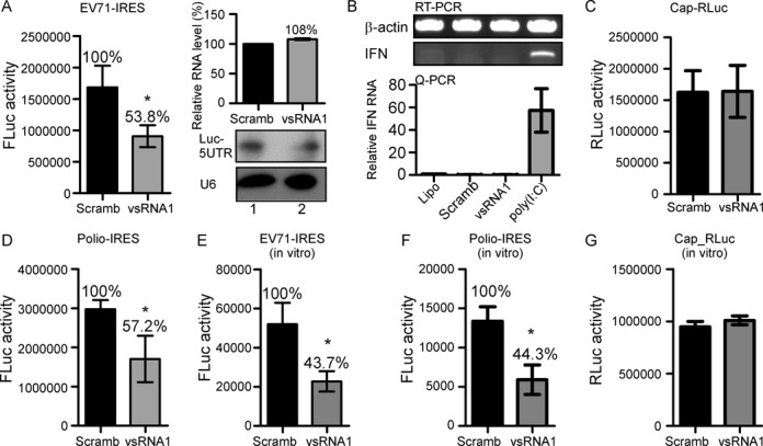
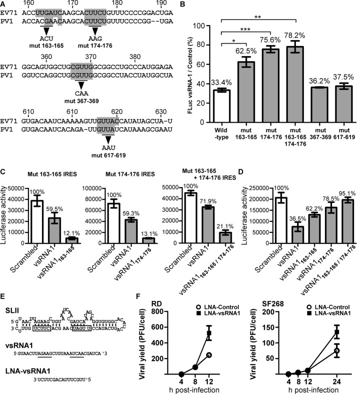
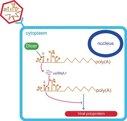
The roles of virus-derived small RNAs (vsRNAs) have been studied in plants and insects. However, the generation and function of small RNAs from cytoplasmic RNA viruses in mammalian cells remain unexplored. This study describes four vsRNAs that were detected in enterovirus 71-infected cells using next-generation sequencing and northern blots. Viral infection produced substantial levels (>10(5) copy numbers per cell) of vsRNA1, one of the four vsRNAs. We also demonstrated that Dicer is involved in vsRNA1 generation in infected cells. vsRNA1 overexpression inhibited viral translation and internal ribosomal entry site (IRES) activity in infected cells. Conversely, blocking vsRNA1 enhanced viral yield and viral protein synthesis. We also present evidence that vsRNA1 targets stem-loop II of the viral 5' untranslated region and inhibits the activity of the IRES through this sequence-specific targeting. Our study demonstrates the ability of a cytoplasmic RNA virus to generate functional vsRNA in mammalian cells. In addition, we also demonstrate a potential novel mechanism for a positive-stranded RNA virus to regulate viral translation: generating a vsRNA that targets the IRES.


Figures

Figure 1.
Figure 1.
Identification of vsRNAs within the EV71 5′UTR. (A) Virus-derived small RNAs (vsRNAs) in SF268 cells infected with EV71 were sequenced using Illumina technology. The position distributions and abundance (reads) of that sequenced vsRNAs that perfectly matched the EV71 5′UTR are shown. (B) vsRNA1–4 (black arrow) in mock- or EV71-infected SF268 cells were detected with 32P-labelled probes containing nucleotides against positions 105–133, 178–198, 522–546 and 694–715 of the EV71 5′UTR. U6 snRNA was used as a loading control. (C) The secondary RNA structure of stem-loop II of the EV71 5′UTR was predicted using Context Fold software (37) and was illustrated with jViz.RNA 2.0 software (http://jviz.cs.sfu.ca/). Circles indicate the predicted vsRNA1 generation site, based on the deep sequencing result. (D) Precursor RNA (upper panel) and vsRNA1 (middle panel) from wild-type EV71 replicon or the vsRNA1 deletion mutant (Δ105–133) replicon EV71 in transfected SF268 cells were detected using 32P-labelled probes against nt 250–270 and nt 105–133 of the viral genome. U6 snRNA was used as a loading control (lower panel). (E) At 3, 6, 9 and 12 h p.i. with EV71, vsRNA1 in human rhabdomyosarcoma (RD) and SF268 cells (from 2.5 × 10⁶ cells per lane) was detected and compared to the indicated levels of synthetic vsRNA1 (mimic vsRNA1). RNA in mock-infected cells (m) served as a negative control. Viral protein 3C and actin were detected by western blotting.
Figure 2.
Figure 2.
Dicer cleaves the EV71 5′UTR into vsRNA1. (A) vsRNA1 in SF268 cells transfected with the control plasmid (shControl) or a plasmid expressing shRNA against Dicer (shDicer) was detected using the vsRNA1 probe. The levels of Dicer and viral 3C protein in these cells were detected by western blotting. (B) In vitro EV71 5′UTR cleavage assay with recombinant Dicer. The 32P -labelled EV71 5′UTR and 3′UTR RNA was detected after being incubated with reaction buffer only (-) or with various amounts (from 0.005 to 0.25 U) of recombinant Dicer for 1.5 h at 37°C. (C) Dicer protein contains 2 RNAse III domains (RIIIDa and RIIIDb) and a dsRBD. N-terminal FLAG-fused Dicer protein (WT) and mutated Dicer proteins with 4 point mutations (*) in the catalytic sites of RIIIDa / RIIIDb (MUT) and with a dsRBD deletion (ΔdsRBD) were used for the in vitro cleavage of the EV71 5′UTR RNA. The 32P-labelled EV71 5′UTR RNA was detected after being incubated with reaction buffer or with these FLAG-Dicer proteins at 37°C for 1.5 h (upper right panel). The FLAG-Dicer proteins in each reaction were detected using an antibody against the FLAG peptide (lower right panel). (D) A vsRNA1 probe was used to detect vsRNA1 generated by EV71-infected cells (Cell RNA) and by synthetic EV71 5′UTR RNA after Dicer treatment (Dicer-treated). RNA isolated from mock-infected cells and from a synthetic 5′UTR without Dicer treatment (Un-treated) served as negative controls. (E) Detection of vsRNA1 in wild-type (WT) or Δ105–133 mutant EV71 replicon RNA treated with recombinant Dicer. Full-length replicon RNA and vsRNA1 were detected using a 32P-labelled probe against nt 250–270 and nt 105–133 of the viral genome.
Figure 3.
Figure 3.
The association between Dicer and the EV71 IRES. (A) In vitro Dicer and Drosha were coprecipitated with EV71 5′UTR. Proteins from SF268 cell lysates were incubated with biotin-UTP (biotin only), unlabelled (Non-bio), or biotin-labelled (Biotin) EV71 5′UTR RNA and streptavidin beads. Proteins from lysates without incubation (No RNA) were pulled down by the same beads and served as a negative control. The Dicer and Drosha proteins in the pull-down reactions or in 1/10 of the SF268 lysate before the pull-down (Input) were detected using western blotting. (B) The same in vitro Dicer and Drosha pull-down assay as (A) was performed on lysates from RD, SK-N-MC and SF268 cells. (C) Competition assay for Dicer association with the EV71 5′UTR. Lane 1 contained the cell lysate (Input) only. The unlabelled EV71 5′UTR (Nonbio-EV71 5′UTR; zero (-) to 30 μg) or poly(A) RNA was added to compete with 3 μg of the biotinylated EV71 5′UTR probe interacting with Dicer in the SF268 cell lysate. Protein that was pulled down without adding any RNA was the negative control (No RNA). (D and E) The complex between recombinant Dicer and the EV71 5′UTR RNA. The 32P-labelled EV71 5′UTR RNA was incubated with 0.4 U of recombinant Dicer (+Dicer) and varying amounts (40 to 400 ng) of unlabelled EV71 5′UTR RNA (+cold 5′UTR) (D) or poly(A) RNA (E). The resulting complexes were analysed using native gel [non-denaturing 3% polyacrylamide TBE gels (29:1 acryamide:bisacryamide)] ectrophoresis. EV71 5′UTR RNA that was not incubated with Dicer and cold 5′UTR (Free 32P 5′UTR) were used to determine the free RNA size. (F) EV71 5′UTR RNA-protein complex formed with varying amounts of recombinant Dicer. Free 32P-labelled EV71 5′UTR RNA only (-) or 5′UTR RNA incubated with varying amounts (0.08 to 0.8 U) of recombinant Dicer (+Dicer) was analysed using the same native gel electrophoresis. (G) The locations of Dicer and Drosha in EV71-infected SF268 cells were detected using specific antibodies at 6 h p.i. The viral 2B and 3D proteins in SF268 cells were used as markers of infection. The nuclei of the cells were stained with Hoechst dye.
Figure 4.
Figure 4.
The effect of vsRNA1 on virus infection and viral protein synthesis. (A) Effect of anti-vsRNA1 sponge RNA on EV71 viral growth. EV71-infected RD cells were transfected with the anti-vsRNA1 sponge or control RNA. After viral adsorption, viruses from the debris and the supernatant were collected at 4, 8 and 12 h p.i. The viral yields were determined with plaque assays. Error bars represent standard deviations for duplicate assays (*P < 0.05; **P < 0.01, Student's t-test). (B) Effect of anti-vsRNA1 sponge RNA on EV71 replication. The luciferase activity in the EV71 replicon in RD cells cotransfected with anti-vsRNA1 sponge or control RNA was monitored at 4, 6, 9, 12 and 15 h post-transfection. Error bars represent standard deviations for three independent experiments (*P < 0.01; ***P < 0.001; Student's t-test). (C) Effects of anti-vsRNA1 sponge on viral and host protein synthesis. Mock- or EV71-infected cells were transfected with the anti-vsRNA1 sponge or control RNA. Protein synthesis in these cells was examined using 35S-methionine/cysteine-labelling between 6 and 7 h p.i. The labelled viral proteins were identified according to their sizes. Viral protein 3C in the cell lysates was detected by western blotting. (D) Effect of vsRNA1 on EV71 viral growth. EV71-infected RD cells were transfected with vsRNA1 mimic or scrambled RNA. After viral adsorption, viruses from the debris and the supernatant were collected at 4, 8 and 12 h p.i. The viral yields were determined using plaque assays. Error bars represent standard deviations for duplicate assays (***P <0.001), Student's t-test). (E) Effect of the vsRNA1 mimic on EV71 replication. Luciferase activity in the EV71 replicon in RD cells cotransfected with vsRNA1 mimic or scrambled RNA was monitored at 4, 6, 9, 12 and 15 h post-transfection. Error bars represent standard deviations for three independent experiments (*P < 0.05, ***P < 0.001, Student's t-test). (F) Effects of vsRNA1 on viral and host protein synthesis. Mock- or EV71-infected cells were transfected with vsRNA1 mimic or scrambled RNA (Scramb). Protein synthesis in these cells was examined using 35S-methionine/cysteine-labelling between 4 and 5 h p.i. The labelled viral proteins were identified according to their sizes. Viral protein 3C in the cell lysates was detected by western blotting.
Figure 5.
Figure 5.
vsRNA1 inhibits viral IRES activity in vitro and in vivo. (A) Effect of vsRNA1 on EV71 IRES activity. SF268 cells were cotransfected with either the vsRNA1 mimic or scrambled RNA (Scramb) combined with a reporter RNA containing the EV71 IRES and the firefly luciferase gene (EV71 IRES-FLuc). The EV71 IRES-driven luciferase expression (FLuc activity) is represented by bars. Error bars represent standard deviations (n = 3, *P < 0.05, Student's t-test). The levels of the transfected reporter RNA (Luc-5′UTR) in the cells cotransfected with scrambled RNA (Scramb) or vsRNA1 were assessed using quantitative real-time RT-PCR (qPCR) (upper right panel) and northern blotting (lower right panel). (B) IFN-β mRNAs in Lipofectamine 2000-treated (Lipo) SF268 cells or cells transfected with scrambled RNA (Scramb); vsRNA1 and poly (I:C) were detected using qPCR. Error bars represent standard deviations (n = 3). (C) The effect of the vsRNA1 mimic on cap-dependent translation using a reporter RNA containing cap and Renilla luciferase (Cap-RLuc). (D) RNAs containing the poliovirus IRES and the firefly luciferase gene (polio IRES-FLuc) were also examined (n = 3, *P < 0.05, Student's t-test). The effects of vsRNA1 on EV71 IRES activity (E), poliovirus IRES activity (F), and cap-dependent translation (G) were also examined in vitro. Reporter RNAs incubated with the vsRNA1 mimic or scrambled RNA (Scramb) were examined in an in vitro translation assay using a mixture of SF268 cell lysate and rabbit reticulocyte lysate (RRL). The IRES- or cap-driven translation activity was determined according to the detected luciferase expression (FLuc activity and RLuc activity). Error bars represent standard deviations for three independent experiments (*P < 0.05, Student's t-test).
Figure 6.
Figure 6.
vsRNA1 target sites on IRES. (A) Predicted vsRNA1 target sites in the EV71 IRES (marked in grey) were selected based on conservation with poliovirus (PV1). Based on the prediction, we generated mutations in nt 163–165, 174–176, 367–369 and 617–619 of the EV71 IRES in the reporter RNA. (B) The inhibitory effects of vsRNA1 on WT and mutant IRES reporter RNAs. Each reporter RNA (including WT, mut 163–165, mut 174–176, mut 163–165, 174–175, mut 367–369 and mut 617–619) was treated with vsRNA1 or scrambled (control) RNA in an in vitro translation assay. The luciferase activity in the vsRNA1 treatment assay was compared to the activity in the scrambled RNA treatment assay, and the percentage of IRES activity under vsRNA1 treatment was calculated separately. Error bars represent standard deviations for three independent experiments (*P < 0.05, **P < 0.01, ***P < 0.001, Student's t-test). (C) Mutant IRES reporter RNAs were treated with the scrambled or vsRNA1 (or modified vsRNA1) that corresponded to each target site mutant (vsRNA1163165 to mut 163–165 IRES, vsRNA1 174–176 to mut 174–176 RNA and vsRNA1 163–165/174–176 to mut 163–165 + 174–176) in the in vitro translation assays. (D) WT IRES reporter RNA was treated with scrambled RNA or vsRNA1 (or modified vsRNA1) in an in vitro translation assay. Error bars represent standard deviations for three independent experiments. (E) LNA against vsRNA1 was generated complementary to the active sites (underlined) on vsRNA1 that target stem-loop II (SLII) of the EV71 5′UTR (the boxes indicate the vsRNA1 target sites). Effect of anti-vsRNA1 LNA on EV71 viral growth. (F) EV71-infected RD and SF268 cells (MOI of 5) were transfected with LNA-vsRNA1 or LNA-control. After viral adsorption, viruses from the debris and the supernatant were collected at the indicated times p.i. The viral yields were determined using plaque assays. Error bars represent standard deviations for duplicated assays.
Figure 7.
Figure 7.
Proposed model for the generation and function of vsRNA1. During EV71 infection, a small proportion of viral RNA is processed by Dicer, and vsRNA1 is generated from the IRES (purple). vsRNA1 targets stem-loop II of the viral IRES (marked in green) and downregulates the translation of viral proteins.

References

    1. Kim V.N., Han J., Siomi M.C. Biogenesis of small RNAs in animals. Nat. Rev. Mol. Cell Biol. 2009;10:126–139. - PubMed
    1. Krol J., Loedige I., Filipowicz W. The widespread regulation of microRNA biogenesis, function and decay. Nat. Rev. Genet. 2010;11:597–610. - PubMed
    1. Siomi M.C., Sato K., Pezic D., Aravin A.A. PIWI-interacting small RNAs: the vanguard of genome defence. Nat. Rev. Mol. Cell Biol. 2011;12:246–258. - PubMed
    1. Cullen B.R. Viruses and microRNAs. Nat. Genet. 2006;38(Suppl):S25–S30. - PubMed
    1. Ding S.W., Voinnet O. Antiviral immunity directed by small RNAs. Cell. 2007;130:413–426. - PMC - PubMed

Publication types