

The first case of Capillaria hepatica infection in a nutria (Myocastor coypus) in Korea
- PMID: 25352702
- PMCID: PMC4210736
- DOI: 10.3347/kjp.2014.52.5.527
The first case of Capillaria hepatica infection in a nutria (Myocastor coypus) in Korea
Abstract
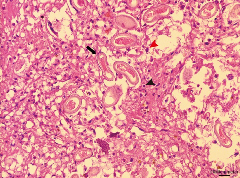
This study reports the first case of Capillaria hepatica infection in a nutria in Korea. Ten nutrias, captured near the Nakdong River, were submitted to our laboratory for necropsy. White-yellowish nodules were found in the liver of 1 of the nutrias at necropsy. Histologically, the lesions were granulomatous, and infiltrations of lipid-laden macrophages, eosinophils, and several multinucleated giant cells were observed. The lesions consisted of numerous eggs and necrotic hepatocytes. The eggs were lemon-shaped and had polar plugs at the ends of both long sides. The eggs were morphologically identified as those of C. hepatica. Worldwide, C. hepatica infection in nutrias is very rare. Nutrias are a kind of livestock, as well as wildlife; therefore, an epidemiological study for parasitic infections needs to be conducted.
Keywords: Capillaria hepatica; liver; nutria.
Conflict of interest statement
We have no conflict of interest related to this study.
Figures


References
-
- Matouch O, Dousek J, Ondracek O. Vyskyt vztekliny u nutrie [Rabies in the nutria] Veterinarstvi. 1978;28:549.
-
- Woods CA, Contreras L, Willner-Chapman G, Whidden HP. Myocastor coypus. Mamm Species. 1992;398:1–8.
-
- Evans J. About nutria and their control. USBSFW, Resource Publication. 1970;86:1–65.
-
- Safarov YB, Kurbanova MA. Influence of some therapeutic substances on the immulogical response of nutria (Myocastor coypus) (antibiotics and nitrofurans in relation to salmonellosis) Veterinariya (Moscow) 1976;11:65–66.
-
- Jelinek P, Valicek L, Smid B, Halouzka R. Demonstration of papillomatosis in nutrias (Myocastor coypus Molina) Vet Med (Praha) 1978;23:113–119. - PubMed
Publication types
MeSH terms
LinkOut - more resources
Full Text Sources
Other Literature Sources
