. 2014 Sep 20:20:1307-17.
eCollection 2014.

Severe retinal degeneration in women with a c.2543del mutation in ORF15 of the RPGR gene

Affiliations

Severe retinal degeneration in women with a c.2543del mutation in ORF15 of the RPGR gene

Bohdan Kousal et al. Mol Vis. .

Abstract

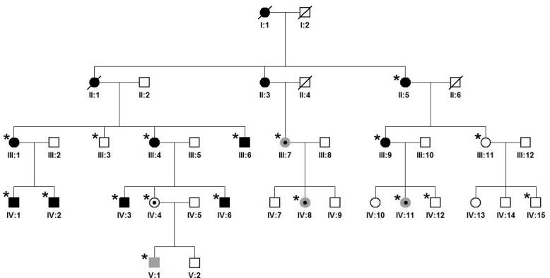
Purpose: To describe the genotype-phenotype correlation and serial observations in a five-generation Czech family with X-linked retinitis pigmentosa (XLRP) associated with severe visual impairment in women.

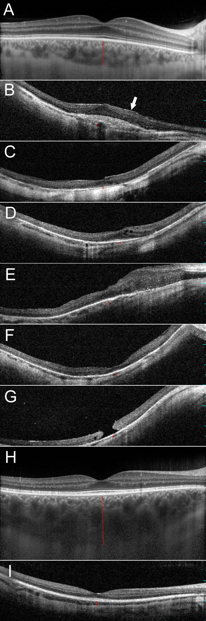
Methods: Comprehensive ophthalmological examination including spectral domain optical coherence tomography (SD-OCT) was performed. Based on the pedigree structure and women being severely affected, autosomal dominant inheritance was suspected, and screening for known mutations by genotyping microarray was performed. Subsequently, direct sequencing of ORF15 RPGR was undertaken.

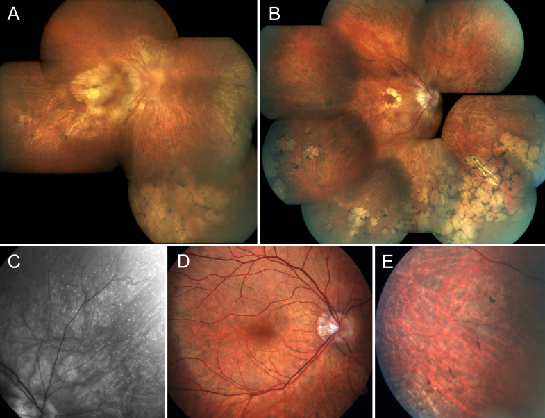
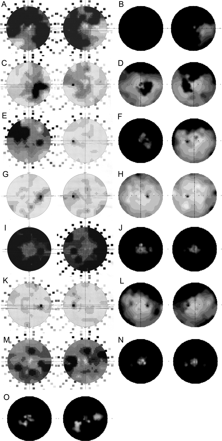
Results: Eighteen family members (nine women and nine men) were examined. A pathogenic variant, c.2543del in ORF15 of RPGR, was found to segregate with disease. The oldest woman and her two sisters had no perception of light in their sixth decade. Four women and five men had signs and symptoms of typical XLRP, including moderate to high myopia. Three other women also had moderate to high myopia and myopic astigmatism but without the presence of bone spicule-like formation. Severe disruption of macular architecture on SD-OCT was equally common in both sexes. Only one 32-year-old female carrier had clinically normal findings. Subfoveal choroidal thickness was decreased in all affected men and in all female carriers, except the only carrier with a normal fundus examination.

Conclusions: The c.2543del mutation in ORF15 of RPGR is associated with a severe phenotype in the women in this family. The presence of a significant myopic refractive error, in the absence of male-to-male transmission, may be indicative of X-linked inheritance. Measurements of choroidal thickness may help in clinically identifying carrier status.


Figures

Figure 1
Figure 1
Pedigree of a family with retinitis pigmentosa associated with a c.2543del mutation in RPGR ORF15. Clinically examined individuals are indicated with an asterisk. All family members with bone spicules indicative of typical retinitis pigmentosa (RP) are shown shaded in black as affected. Individuals with myopia (−3.0 to −15.0 diopter sphere) but no pigmentary retinopathy are shown shaded in gray. Individuals III:4, III:9, IV:2, IV:4, and IV:11 were positive for the disease-causing mutation, whereas III:3 and IV:12 were negative. For individuals II:5, III:1, III:6, III:7, III:11, IV:1, IV:3, IV:6, IV:8, IV:15, and V:1, no DNA was available for laboratory investigation.
Figure 2
Figure 2
Spectrum of fundus finding in two women and one man with retinitis pigmentosa caused by a c.2543del RPGR ORF15 mutation. A: Composite fundus photography of the right eye of female III:4 at the age of 54 years. B: Composite fundus photography of the right eye of female III:9 at the age of 50 years; note the chorioretinal degeneration at the macula and in the periphery with intraretinal asymmetric involvement. C: Red-free imaging enhancing peripheral white dots in the upper nasal quadrant of the right eye of female III:9. D, E: Fundus photographs of male IV:6 aged 19; note the relatively normal appearing macula and pigment clumps in the mid-periphery.
Figure 3
Figure 3
Horizontal optical coherence tomography images cross sectioning the fovea in the right eyes of affected men and women who carry a c.2543del mutation in RPGR ORF15. Choroidal thickness is indicated with a red bar. A: Normal retinal architecture in a 40-year-old control individual (enhanced depth imaging). B: Chorioretinal thinning, missing foveal depression, scar formation, and epiretinal membrane (arrow) in female III:1 at the age of 64 years. C: Chorioretinal thinning, missing foveal depression, and vitreomacular traction associated with foveal elevation in female III:4 at the age of 54 years. D: Chorioretinal thinning, myopic foveoschisis, and posterior staphyloma in female III:9 at the age of 50 years. E: Chorioretinal thinning and focal retinal thickening in male IV:1 at the age of 42 years. F: Chorioretinal thinning and myopic posterior staphyloma in male IV:2 at the age of 38 years. G: Chorioretinal thinning, macular hole, and myopic posterior staphyloma in male IV:3 at the age of 30 years. H: Normal retinal and choroidal architecture in female IV:4 at the age of 32 years (enhanced depth imaging). I: Choroidal thinning and normal retinal architecture in male IV:6 at the age of 19 years.
Figure 4
Figure 4
Serial observations of disease progression on automated visual field for individuals with a c.2543del RPGR ORF15 mutation. Visual field scans (showing 50° nasal and temporal to fixation) of the right and left eyes of female III:4 at the age of 47 (A) and 58 (B) years; the right and left eyes of female III:1 at the age of 58 (C) and 64 (D) years; the right and left eyes of female III:9 at the age of 44 (E) and 50 (F) years; the right and left eyes of female IV:4 at the age of 25 (G) and 29 (H) years; the right and left eyes of male IV:3 at the age of 23 (I) and 30 (J) years; the right and left eyes of male IV:6 at the age of 14 (K) and 19 (L) years; the right and left eyes of male IV:2 at the age of 32 (M) and 38 (N) years; the right and left eyes of male IV:1 at the age of 42 (O) years. Static perimetry results are shown as 12-level gray scales of sensitivity loss. Black areas indicate no detection of stimuli. Physiologic blind spot is shown as a black circle at 12° in the temporal field.

References

    1. Daiger SP, Sullivan LS, Bowne SJ. Genes and mutations causing retinitis pigmentosa. Clin Genet. 2013;84:132–41. - PMC - PubMed
    1. Berger W, Kloeckener-Gruissem B, Neidhardt J. The molecular basis of human retinal and vitreoretinal diseases. Prog Retin Eye Res. 2010;29:335–75. - PubMed
    1. Hartong DT, Berson EL, Dryja TP. Retinitis pigmentosa. Lancet. 2006;368:1795–809. - PubMed
    1. Koenekoop RK, Loyer M, Hand CK, Al Mahdi H, Dembinska O, Beneish R, Racine J, Rouleau GA. Novel RPGR mutations with distinct retinitis pigmentosa phenotypes in French-Canadian families. Am J Ophthalmol. 2003;136:678–87. - PubMed
    1. Banin E, Mizrahi-Meissonnier L, Neis R, Silverstein S, Magyar I, Abeliovich D, Roepman R, Berger W, Rosenberg T, Sharon D. A non-ancestral RPGR missense mutation in families with either recessive or semi-dominant X-linked retinitis pigmentosa. Am J Med Genet A. 2007;143A:1150–8. - PubMed

Publication types

MeSH terms

LinkOut - more resources