Review
. 2014 Oct 13:8:114.
doi: 10.3389/fnana.2014.00114. eCollection 2014.

Neuronal MHC-I expression and its implications in synaptic function, axonal regeneration and Parkinson's and other brain diseases

Affiliations
Review

Neuronal MHC-I expression and its implications in synaptic function, axonal regeneration and Parkinson's and other brain diseases

Carolina Cebrián et al. Front Neuroanat. .

Abstract

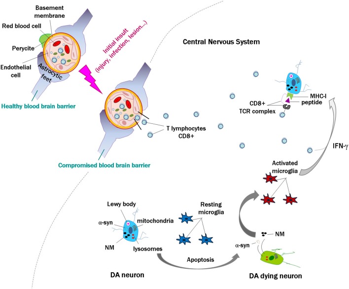
Neuronal expression of major histocompatibility complex I (MHC-I) has been implicated in developmental synaptic plasticity and axonal regeneration in the central nervous system (CNS), but recent findings demonstrate that constitutive neuronal MHC-I can also be involved in neurodegenerative diseases by playing a neuroinflammtory role. Recent reports demonstrate its expression in vitro and in human postmortem samples and support a role in neurodegeneration involving proinflammatory cytokines, activated microglia and increased cytosolic oxidative stress. Major histocompatibility complex I may be important for both normal development and pathogenesis of some CNS diseases including Parkinson's.

Keywords: major histocompatibility complex class I; neurodegeneration; neuroinflammation; neurons; plasticity.


Figures

Figure 1
Figure 1
Model of a CD8+ cytotoxic T cell targeting an MHC-I expressing cell. Intracellular antigens are digested by the proteasome into small peptides. A specialized carrier, the transporter associated with antigen processing (TAP) complex, translocates the peptide into the endoplasmic reticulum (ER), allowing the antigen to bind MHC class I, which consists of an alpha and beta 2 microglobulin (β2m) chain. Endoplasmic reticulum-derived vesicles that contain the complex fuse with the Golgi. The complex is then packaged in secretory vesicles that fuse with the plasma membrane to insert the complex on the cell surface. Major histocompatibility complex I molecules present antigens to CD8+ cytotoxic T cells. The T cell CD8+ molecule recognizes MHC-I, while TCRs recognize specific antigenic peptides. The TCR complex contains CD3 and ζ(zeta) chains. Once the CD8+ T cell recognizes the target cell, apoptosis can occur in two ways: one uses the Fas ligand protein, which is expressed on the surface of the CD8+ T cells and binds to the Fas receptor on the target cell, which triggers apoptosis through the classical caspase cascade. The other uses secretion of granzyme B and perforin from T cell granules into the intercellular space between the cells.
Figure 2
Figure 2
Scheme modeling an initial insult and disruption of the blood brain barrier, with penetration of lymphocytes into the brain. In the event of death of a few catecholamine neurons, alpha-synuclein (α-syn) and neuromelanin (NM) are released to the extracellular space, activating microglial cells that in turn release proinflammatory substances such as interferon gamma (IFN-γ). This would induce major histocompatibility class I expression (MHC-I) in the membrane of catecholamine neurons, and if they contain proteins that are misfolded or cannot be normally degraded, these might be presented as antigens by MHC-I. Cytotoxic lymphocytes that enter the brain may recognize these presented antigens and kill the neurons, which would again release α-syn and NM. This could continue a vicious cycle that may contribute to the death of catecholamine neurons in Parkinson’s disease.

References

    1. Abraham S., Nagaraj A. S., Basak S., Manjunath R. (2010). Japanese encephalitis virus utilizes the canonical pathway to activate NF-kappaB but it utilizes the type I interferon pathway to induce major histocompatibility complex class I expression in mouse embryonic fibroblasts. J. Virol. 84, 5485–5493 10.1128/JVI.02250-09 - DOI - PMC - PubMed
    1. Almeida C. M., Galvão Mde L., Ferreira P. L., Braga W. S. (2009). Interferon-induced Parkinsonism in a patient with chronic hepatitis C. Arq. Neuropsiquiatr. 67, 715–716 10.1590/s0004-282x2009000400031 - DOI - PubMed
    1. Andersen M. H., Schrama D., Thor Straten P., Becker J. C. (2006). Cytotoxic T cells. J. Invest. Dermatol. 126, 32–41 10.1038/sj.jid.5700001 - DOI - PubMed
    1. Barrow A. D., Trowsdale J. (2008). The extended human leukocyte receptor complex: diverse ways of modulating immune responses. Immunol. Rev. 224, 98–123 10.1111/j.1600-065X.2008.00653.x - DOI - PubMed
    1. Béraud D., Hathaway H. A., Trecki J., Chasovskikh S., Johnson D. A., Johnson J. A., et al. (2013). Microglial activation and antioxidant responses induced by the Parkinson’s disease protein α-synuclein. J. Neuroimmune Pharmacol. 8, 94–117 10.1007/s11481-012-9401-0 - DOI - PMC - PubMed