

Solitary fibrous tumour of the pleura - cases analysis
- PMID: 25352940
- PMCID: PMC4210359
- DOI: 10.12659/PJR.890525
Solitary fibrous tumour of the pleura - cases analysis
Abstract
Background: Isolated fibrous tumor of the pleura (SFTP - Solitary fibrous tumour of the pleura/localized fibrous tumour of the pleura) is a rare primary tumour of the pleura of mesenchymal origin. In most cases, it is a benign lesion. It is composed of spindle cells similar to fibroblasts and derives probably from submesothelial mesenchyme. The aim of the study was to analyze clinical symptoms, incidence, possibility of suggesting the diagnosis on the basis of imaging tests, and confirmation of the diagnosis in pathological tests with regard to studies of histochemistry examination.
Material/methods: Clinical and morphological material obtained from 14 patients from Department of Thoracic Surgery of Subcarpathian Chest Disease Center treated between year 2004 and 2010 was analysed. In the first stage, selected cases of patients with isolated fibrous tumour of the pleura were chosen from the archives and the analysis of their medical history was carried out. Basic information about age, gender, medical history, smoking habit, physical examination and results of imaging, endoscopic and morphological examinations were noted. The second parallel component of the study was pathomorphological examinations of the surgical material obtained from the patients, including the assessment of morphology and immunohistochemistry.
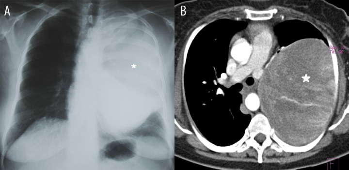
Results: Of the 14 examined patients, fibrous tumour occurred in 8 men and 6 women. The age range of the patients was 37-73 years, with a peak attributable to the 6(th) decade of life. In 8 patients the tumour was detected incidentally during routine examinations. In 7 patients there were no clinical signs of respiratory disease, and if present, then the most common complaint was shortness of breath. Regarding symptoms not connected with the respiratory system, anemia occurred most frequently. Fibrous tumour of the pleura was more often associated with the visceral pleura than with the parietal pleura. The largest lesion was approximately 20 cm in size.
Conclusions: Fibrous tumour of the pleura is a pleura-based neoplasm which is usually detected incidentally, and is often asymptomatic or poorly symptomatic. Computed tomography imaging allows to suggest a correct diagnosis. Histopathological diagnosis is based on immunohistochemical examinations.
Keywords: Immunohistochemistry; Pleural –blood; Solitary Fibrous Tumor; Spiral Computed; Tomography.
Figures


References
-
- Masaya O, Hiroyasu Y, Sung-Soo Ch, et al. Solitary fibrous tumor of the pleura presenting dry cough induced by postural posittion. Ann Thorac Cardiovasc Surg. 2009;15(6):401–3. - PubMed
-
- Robinson LA. Solitary fibrous tumor of the pleura. Cancel Control, Journal of the Moffiti Cancer Center. 2006;13(4):264–69. - PubMed
-
- Pinedo-Onofre JA, Robles-Perez A, Pena-Mirabal ES, et al. Giant solitary fibrous tumor of the pleura. Cir Cir. 2010;78(1):31–42. - PubMed
-
- Findik G, Ozturk F, Gunay E, et al. Surgical management of solitary fibrous tumors of the pleura – an analysis of 21 cases. Adv Clin Exp Med. 2011;20(3):363–69.
LinkOut - more resources
Full Text Sources
Other Literature Sources