. 2015 Jan;95(1):64-79.
doi: 10.1111/mmi.12841. Epub 2014 Nov 24.

Genome-scale quantitative characterization of bacterial protein localization dynamics throughout the cell cycle

Affiliations
Free PMC article

Genome-scale quantitative characterization of bacterial protein localization dynamics throughout the cell cycle

Nathan J Kuwada et al. Mol Microbiol. 2015 Jan.
Free PMC article

Abstract

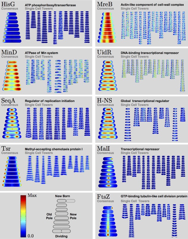
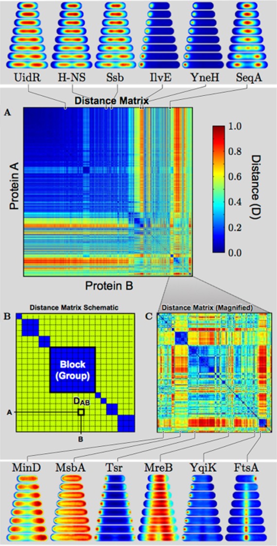
Bacterial cells display both spatial and temporal organization, and this complex structure is known to play a central role in cellular function. Although nearly one-fifth of all proteins in Escherichia coli localize to specific subcellular locations, fundamental questions remain about how cellular-scale structure is encoded at the level of molecular-scale interactions. One significant limitation to our understanding is that the localization behavior of only a small subset of proteins has been characterized in detail. As an essential step toward a global model of protein localization in bacteria, we capture and quantitatively analyze spatial and temporal protein localization patterns throughout the cell cycle for nearly every protein in E. coli that exhibits nondiffuse localization. This genome-scale analysis reveals significant complexity in patterning, notably in the behavior of DNA-binding proteins. Complete cell-cycle imaging also facilitates analysis of protein partitioning to daughter cells at division, revealing a broad and robust assortment of asymmetric partitioning behaviors.


Figures

Fig. 1
Fig. 1
Consensus localization patterns and single-cell tower images for proteins HisG, MreB, MinD, UidR, SeqA, H-NS, Tsr, MalI and FtsZ. Single-cell tower images capture protein localization dynamics in single cells. For each protein, 9–15 complete cell cycles are shown in false color. For SeqA, like many proteins in the collection, the single-cell tower images display significant cell-to-cell variation in protein localization, despite qualitative similarities. To visualize average dynamics and to facilitate the quantitative comparison between protein localization patterns, we compute the consensus localization pattern by computing the mean localization pattern over all single-cell data for each protein.
Fig. 2
Fig. 2
Cell-cycle timing probed by consensus localization patterns. To demonstrate the ability of consensus localization patterns to determine cell-cycle timing, we analyze the localization patterns of proteins in two processes where the cell-cycle ordering of protein localization is already known. Consensus location patterns are represented as kymographs. A. SeqA and MukB are proteins implicated in chromosome replication and segregation. Consistent with previously reported results, the kymographs show MukB arriving at the quarter-cell position prior to SeqA. B. FtsZ, MinD and SlmA are proteins implicated in cytokinesis. Both MinD and SlmA are known to inhibit FtsZ-dependent Z-ring formation. The kymographs show significant FtsZ localization at midcell only after MinD and SlmA are depleted from midcell, consistent with the known mechanism of z-ring regulation.
Fig. 3
Fig. 3
Virtual co-localization of all protein pairs measured by distance between consensus localization patterns. (A) The distance matrix is visualized as a heat map with (B) schematic representation of block diagonal form and (C) a magnified region of the distance matrix. Consensus localization patterns that are identical have a distance of zero (dark blue, see colorbar). Representative examples of consensus localization patterns and their positions in the distance matrix are shown above and below the matrix.
Fig. 4
Fig. 4
Consensus localization pattern diversity analyzed by PCA. A. Schematic view of PCA of consensus localization patterns. In PCA, each consensus localization pattern A is represented as a sum PCi with projection coefficients Ai. The PCs are ordered by their significance (power) in representing the library of consensus localization patterns. The PC with the highest power is labeled PC1. B. Visualization of the first 20 PCs. The PCs should not be interpreted as localization patterns, but rather as the redistribution of protein from the mean consensus localization pattern. C. The power spectrum for the first 200 PCs. There are 17 PCs with power greater than the power corresponding to a single consensus localization pattern.
Fig. 5
Fig. 5
Identification of common localization patterns for DNA-binding proteins. A. Schematic view of PC representation of a consensus localization pattern A as projection coefficients (Ai) of the DNA-binding protein PCD. The PCDs are labeled by their qualitative effect on protein localization, e.g. the ‘Middle’ redistributes protein toward the center of the nucleoid with respect to the cell long axis, while ‘Surface’ rearranges protein to the outer surface of the nucleoid with respect to the short axis of the cell. B. To visualize the effect of each PCD on protein localization, we generated representative patterns shown on each axis of C and D. C. Projections of consensus localization patterns for along PCD1 (‘Midcell’) and PCD2 (‘Ter’). Outlying patterns are labeled by gene name. D. Projections of consensus localization patterns for PCD3 (‘Surface’) and PCD4 (‘Origin’). Outlying patterns are labeled by gene name.
Fig. 6
Fig. 6
Asymmetric protein partitioning at cell division. A. Representative consensus localization patterns and single-cell tower images illustrating symmetric protein partitioning (UidR), enrichment to the old-daughter cell (HisG) and enrichment to the new-daughter cell (MalI). B. Scatter plot of integrated intensity of the new-daughter and old-daughter cells immediately following division for all single-cell data of UidR, HisG and MalI. C. The mean new-daughter (Inew) and mean old-daughter (Iold) integrated intensity for all proteins in the localization library, where green dots represent symmetric partitioning between daughter cells, red dots represent enrichment to the old-daughter cell and blue dots represent enrichment to the new-daughter cell. For reference, the positions of UidR, HisG and MalI are indicated.
Fig. 7
Fig. 7
Diversity in polar localization timing. Representative consensus localization patterns for proteins that localize to the cell poles reveal a wide range of localization timing. Arrows indicate the qualitative arrival time of proteins to the new cell pole. Proteins like PerR, ZapA, TolQ and KdtA arrive at midcell (the nacent new pole) prior to division, but with significantly different arrival times, while YgeD, Tap, WcaB arrive at the new cell pole well into the following cell cycle. Proteins such as SelA appear to never significantly accumulate at the new cell pole.

References

    1. Alley MR, Maddock JR. Shapiro L. Polar localization of a bacterial chemoreceptor. Genes Dev. 1992;6:825–836. - PubMed
    1. Baneyx F. Mujacic M. Recombinant protein folding and misfolding in Escherichia coli. Nat Biotechnol. 2004;22:1399–1408. - PubMed
    1. Bendezu FO, Hale CA, Bernhardt TG. de Boer PA. RodZ (YfgA) is required for proper assembly of the MreB actin cytoskeleton and cell shape in E. coli. EMBO J. 2008;28:193–204. - PMC - PubMed
    1. Bernhardt TG. de Boer PA. SlmA, a nucleoid-associated, FtsZ binding protein required for blocking septal ring assembly over chromosomes in E. coli. Mol Cell. 2005;18:555–564. - PMC - PubMed
    1. Bobola N, Jansen RP, Shin TH. Nasmyth K. Asymmetric accumulation of Ash1p in postanaphase nuclei depends on a myosin and restricts yeast mating-type switching to mother cells. Cell. 1996;84:699–709. - PubMed

Publication types

MeSH terms

Substances

LinkOut - more resources