

PDCD10 gene mutations in multiple cerebral cavernous malformations
- PMID: 25354366
- PMCID: PMC4212902
- DOI: 10.1371/journal.pone.0110438
PDCD10 gene mutations in multiple cerebral cavernous malformations
Erratum in
-
Correction: PDCD10 gene mutations in multiple cerebral cavernous malformations.PLoS One. 2015 Apr 7;10(4):e0123486. doi: 10.1371/journal.pone.0123486. eCollection 2015. PLoS One. 2015. PMID: 25849884 Free PMC article. No abstract available.
Abstract
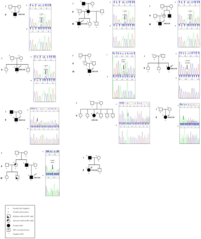
Cerebral cavernous malformations (CCMs) are vascular abnormalities that may cause seizures, intracerebral haemorrhages, and focal neurological deficits. Familial form shows an autosomal dominant pattern of inheritance with incomplete penetrance and variable clinical expression. Three genes have been identified causing familial CCM: KRIT1/CCM1, MGC4607/CCM2, and PDCD10/CCM3. Aim of this study is to report additional PDCD10/CCM3 families poorly described so far which account for 10-15% of hereditary cerebral cavernous malformations. Our group investigated 87 consecutive Italian affected individuals (i.e. positive Magnetic Resonance Imaging) with multiple/familial CCM through direct sequencing and Multiplex Ligation-Dependent Probe Amplification (MLPA) analysis. We identified mutations in over 97.7% of cases, and PDCD10/CCM3 accounts for 13.1%. PDCD10/CCM3 molecular screening revealed four already known mutations and four novel ones. The mutated patients show an earlier onset of clinical manifestations as compared to CCM1/CCM2 mutated patients. The study of further families carrying mutations in PDCD10/CCM3 may help define a possible correlation between genotype and phenotype; an accurate clinical follow up of the subjects would help define more precisely whether mutations in PDCD10/CCM3 lead to a characteristic phenotype.
Conflict of interest statement
Figures


References
-
- Gomori JM, Grossman RI, Goldberg HI, Hackney DB, Zimmerman RA, et al. (1986) Occult cerebral vascular malformations: high-field MR imaging. Radiology 158: 707–713. - PubMed
-
- Labauge P, Denier C, Bergametti F, Tournier-Lasserve E (2007) Genetics of cavernous angiomas. Lancet Neurol 6: 237–244. - PubMed
-
- Riant F, Bergametti F, Fournier H-D, Chapon F, Michalak-Provost S, et al. (2013) CCM3 Mutations Are Associated with Early-Onset Cerebral Hemorrhage and Multiple Meningiomas. Mol Syndromol 4: 165–172 Available: http://www.pubmedcentral.nih.gov/articlerender.fcgi?artid=3666455&tool=p.... Accessed 21 July 2014 - PMC - PubMed
Publication types
MeSH terms
Substances
LinkOut - more resources
Full Text Sources
Other Literature Sources
