. 2014 Oct 30:5:5352.
doi: 10.1038/ncomms6352.

A system for the continuous directed evolution of proteases rapidly reveals drug-resistance mutations

Affiliations

A system for the continuous directed evolution of proteases rapidly reveals drug-resistance mutations

Bryan C Dickinson et al. Nat Commun. .

Abstract

The laboratory evolution of protease enzymes has the potential to generate proteases with therapeutically relevant specificities and to assess the vulnerability of protease inhibitor drug candidates to the evolution of drug resistance. Here we describe a system for the continuous directed evolution of proteases using phage-assisted continuous evolution (PACE) that links the proteolysis of a target peptide to phage propagation through a protease-activated RNA polymerase (PA-RNAP). We use protease PACE in the presence of danoprevir or asunaprevir, two hepatitis C virus (HCV) protease inhibitor drug candidates in clinical trials, to continuously evolve HCV protease variants that exhibit up to 30-fold drug resistance in only 1 to 3 days of PACE. The predominant mutations evolved during PACE are mutations observed to arise in human patients treated with danoprevir or asunaprevir, demonstrating that protease PACE can rapidly identify the vulnerabilities of drug candidates to the evolution of clinically relevant drug resistance.


Figures

Figure 1
Figure 1. Development of a system to link protease activity to gene expression
(a) Protease-activated RNA polymerase (PA-RNAP). T7 RNAP is fused to the natural inhibitor T7 lysozyme through a linker containing a protease target substrate sequence. While the linker is intact, the complex preferentially adopts the lysozyme-bound, RNAP-inactive state. Proteolysis of the target sequence favors dissociation of the complex, freeing active T7 RNAP to transcribe genes downstream of the T7 promoter. This study used an accessory plasmid (AP) in which the T7 promoter drives a tandem gIII-luciferase (lux) cassette. (b) Sequences of the protein linkers containing a target protease substrate used for each PA-RNAP, with T7 lysozyme residues in blue, protease substrates in red, T7 RNAP residues in green, and linker regions in black. (c) Plasmids used for protease PACE. An AP that has gIII and luciferase (lux) under the control of the T7 promoter serves as the source of gIII in the cells. A complementary plasmid (CP) constitutively expresses a PA-RNAP variant with a protease target substrate sequence embedded in the linker. (d) PA-RNAP gene expression response in E. coli cells. Host cells were transformed with (i) an AP containing the T7 promoter driving gIII-lux; (ii) a CP that constitutively expresses a PA-RNAP including the TEV protease substrate, the HCV protease substrate, or the HRV protease substrate; and (iii) a plasmid that expresses TEV protease (orange bars), HCV protease (purple bars), or HRV protease (gray bars). Gene expression is activated only when the expressed protease cleaves the amino acid sequence on the PA-RNAP sensor. The luminescence experiment was performed in triplicate with error bars indicating the standard deviation.
Figure 2
Figure 2. PA-RNAPs link protease activity to phage propagation
(a) The protease PACE system. Fixed volume vessels (lagoons) contain phage in which gIII is replaced with a gene encoding an evolving protease. The lagoon is fed with host cells that contain an AP with the T7 promoter driving gIII and a CP that expresses a PA-RNAP. Phage infect incoming cells and inject their genome containing a protease variant. Only if the protease variant can activate the PA-RNAP by cleaving the linker encoding the target protease substrate, gIII is expressed and that SP can propagate. (b–d) Enrichment of active proteases from mixed populations using PACE. At time 0, a lagoon was seeded with a 1,000-fold excess of non-cognate protease-encoding phage over cognate protease-encoding phage. The lagoon was continuously diluted with host cells containing a PA-RNAP with either the HCV (b), TEV (c), or HRV (d) protease substrates. Lagoon samples were periodically analyzed by PCR. In all three cases, phage encoding the cognate protease were rapidly enriched in the lagoon while phage encoding the non-cognate protease were depleted. Full gels are shown in Supplementary Figure 8.
Figure 3
Figure 3. HCV PA-RNAP response to protease inhibitors in E. coli cells
Host cells expressing the HCV PA-RNAP were incubated with HCV protease inhibitors danoprevir (a) or asunaprevir (b) for 90 min, followed by inoculation with HCV protease encoding phage. After 3 h, luminescence assays were used to quantify relative gene activation resulting from the PA-RNAP. Luminescence experiments were performed in triplicate with error bars depicting the standard deviation.
Figure 4
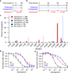
Figure 4. Continuous evolution of drug resistance in HCV protease
(a, b) PACE condition timeline for evolution in the presence of danoprevir (a) or asunaprevir (b). The blue arrows indicate arabinose-induced enhanced mutagenesis, and the red arrow shows the timing and dosing of HCV protease inhibitors. (c) High-throughput sequencing data from phage populations in replicate lagoons (L1 and L2) subjected to danoprevir treatment at 28 h, asunaprevir treatment at 75 h, and no drug at 72 h. All mutations with frequencies more than 1% above the allele-specific error rate are shown. (d) In vitro analysis of danoprevir inhibition of mutant HCV proteases that evolved during PACE. (e) In vitro analysis of asunaprevir inhibition of mutant HCV proteases that evolved during PACE. For (d) and (e), evolved HCV protease variants were expressed and purified, then assayed using an internally quenched fluorescent-substrate (Anaspec). In vitro analyses were performed in triplicate with error bars calculated as the standard deviation.

References

    1. Schilling O, Overall CM. Proteome-derived, database-searchable peptide libraries for identifying protease cleavage sites. Nat Biotechnol. 2008;26:685–694. - PubMed
    1. Walsh G. Biopharmaceutical benchmarks 2006. Nat Biotechnol. 2006;24:769–776. - PubMed
    1. Wehr MC, et al. Monitoring regulated protein-protein interactions using split TEV. Nat Methods. 2006;3:985–993. - PubMed
    1. Craik CS, Page MJ, Madison EL. Proteases as therapeutics. Biochem J. 2011;435:1–16. - PMC - PubMed
    1. Gray DC, Mahrus S, Wells JA. Activation of specific apoptotic caspases with an engineered small-molecule-activated protease. Cell. 2010;142:637–646. - PMC - PubMed

Publication types