. 2014 Oct 29;34(44):14803-15.
doi: 10.1523/JNEUROSCI.2907-14.2014.

Transcranial magnetic stimulation of the prefrontal cortex in awake nonhuman primates evokes a polysynaptic neck muscle response that reflects oculomotor activity at the time of stimulation

Affiliations

Transcranial magnetic stimulation of the prefrontal cortex in awake nonhuman primates evokes a polysynaptic neck muscle response that reflects oculomotor activity at the time of stimulation

Chao Gu et al. J Neurosci. .

Abstract

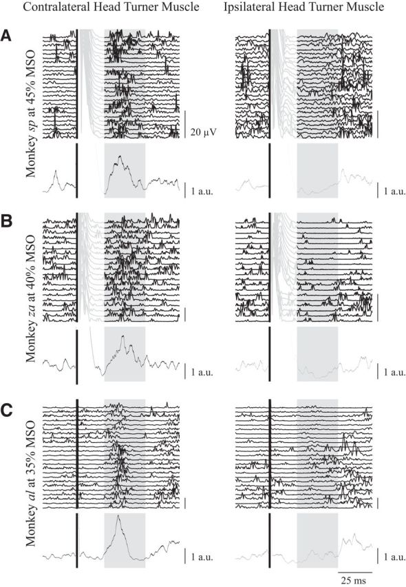
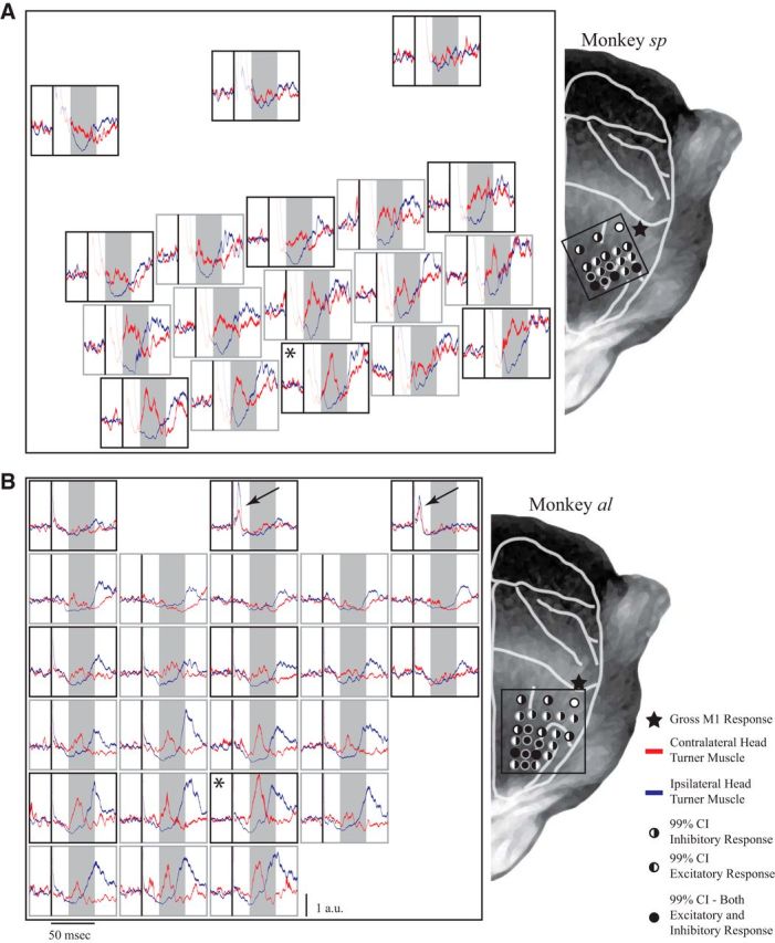
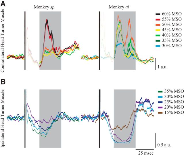
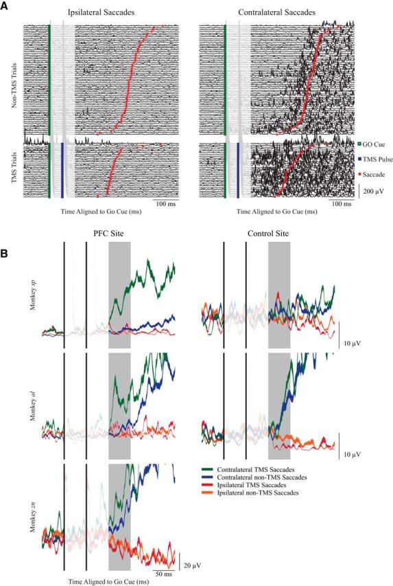
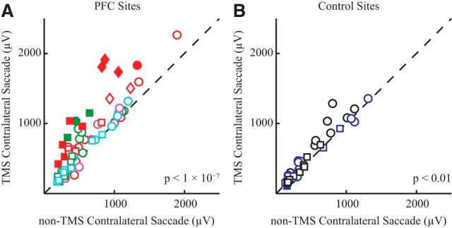
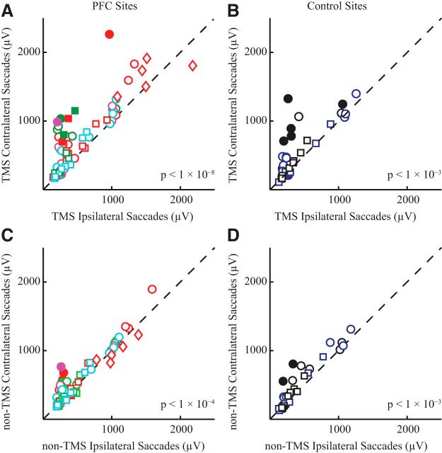
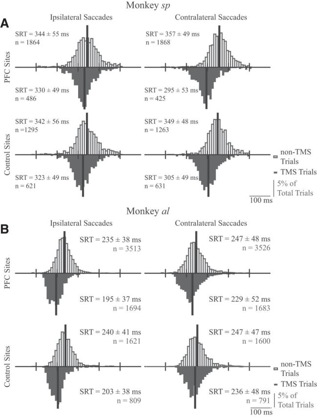
Transcranial magnetic stimulation (TMS) has emerged as an important technique in cognitive neuroscience, permitting causal inferences about the contribution of a given brain area to behavior. Despite widespread use, exactly how TMS influences neural activity throughout an interconnected network, and how such influences ultimately change behavior, remain unclear. The oculomotor system of nonhuman primates (NHPs) offers a potential animal model to bridge this gap. Here, based on results suggesting that neck muscle activity provides a sensitive indicator of oculomotor activation, we show that single pulses of TMS over the frontal eye fields (FEFs) in awake NHPs evoked rapid (within ∼25 ms) and fairly consistent (∼50-75% of all trials) expression of a contralateral head-turning synergy. This neck muscle response resembled that evoked by subsaccadic electrical microstimulation of the FEF. Systematic variation in TMS location revealed that this response could also be evoked from the dorsolateral prefrontal cortex (dlPFC). Combining TMS with an oculomotor task revealed state dependency, with TMS evoking larger neck muscle responses when the stimulated area was actively engaged. Together, these results advance the suitability of the NHP oculomotor system as an animal model for TMS. The polysynaptic neck muscle response evoked by TMS of the prefrontal cortex is a quantifiable trial-by-trial reflection of oculomotor activation, comparable to the monosynaptic motor-evoked potential evoked by TMS of primary motor cortex. Our results also speak to a role for both the FEF and dlPFC in head orienting, presumably via subcortical connections with the superior colliculus.

Keywords: TMS; animal model; frontal cortex; frontal eye fields; oculomotor system; saccades.


Figures

Figure 1.
Figure 1.
A, The locations of the fiducial markers for both monkeys sp and al overlaid on their anatomical MRI scans, with the central and arcuate sulcus highlighted (left subplots), and a representative head of a monkey with the estimated locations of the fiducial makers (right subplots). B, Schematic line drawings of the muscles of interest, which were implanted bilaterally. All three of these muscles contribute to horizontal head turns to the ipsilateral side. C, The memory-guided saccade paradigm. The FP remained illuminated prior, during, and after a peripheral target was flashed 20° left or right. Two TMS pulses (20 Hz, 50 ms apart) were delivered on one-third of all trials, concurrently with FP offset, which also served as the GO cue for the monkey to look at the remembered location of the target.
Figure 2.
Figure 2.
Representative examples from monkey sp (A), monkey zn (B), and monkey al (C) of the contralateral head-turning synergy evoked by single pulses of TMS over the superior arm of the arcuate. Rectified EMG activity aligned to TMS onset (solid black line) is shown for ∼25 trials for contralateral and ipsilateral neck muscles (left and right columns respectively, the last trial is the topmost trace), as well as the session average (bottom lines showing mean ± SE; black or gray traces for contralateral or ipsilateral muscles, respectively), which is normalized to the baseline EMG activity averaged from the 50 ms before TMS onset. In all monkeys, TMS evoked a simultaneous increase or decrease in activity for contralateral or ipsilateral neck muscles ∼20–50 ms after TMS (shaded box). TMS evoked a substantial artifact in monkeys sp and zn, and is shown as recorded in the individual traces in the lighter portions of the lines. Note that this artifact did not contaminate the response window. Scale bar for mean traces show the average baseline activity 50 ms before TMS.
Figure 3.
Figure 3.
Results of the mapping experiment for monkey sp (A) and monkey al (B). Left, Each subplot shows the mean ± SE of contralateral (red) and ipsilateral (blue) neck muscle activity aligned to TMS (black line), normalized to baseline activity at each site; shaded boxes show the response window, as in Figure 2. Right, The depicted sites represent approximate locations of markers projected onto a representation of a monkey's head for both monkeys sp and al. The asterisks in the left panel mark the location associated with the largest contralateral neck muscle response. Black arrows in B show locations from where TMS evoked a distinct profile of bilateral cocontraction in monkey al. Right, The filling of the circles indicates responses that went above or below the 99% CIs calculated from the baseline activity for contralateral and ipsilateral muscles, respectively (see legend). Black stars mark the locations from where a gross twitch from the contralateral hand could be evoked with TMS.
Figure 4.
Figure 4.
The intensity experiment. A, Contralateral neck muscle responses aligned to a single TMS pulse while the NHPs looked at a central FP, scaled for different TMS intensities (same format as Fig. 2). B, Ipsilateral neck muscle responses aligned to a single pulse of TMS while the NHPs looked at an FP 20° ipsilateral to the side of TMS, which increases background activity on the muscle. Note the difference in the TMS intensity needed to evoke an excitatory response on the contralateral neck muscle versus an inhibitory response on the ipsilateral muscle (40% MSO vs 25% MSO for monkey sp and 30% MSO vs 15% MSO for monkey al).
Figure 5.
Figure 5.
State-dependent recruitment of contralateral turning neck muscles in the memory-guided saccade task. A, Neck EMG activity from individual trials from one session of the memory-guided saccade task from monkey sp, aligned to the GO cue (green square). Trials are segregated by saccade direction, and whether TMS was delivered (bottom; the second of the two TMS pulses is shown in the blue square). Trials are ordered by SRT (red circle). B, Representative examples of mean ± SE. EMG activity from contralateral turning muscles from single sessions, with TMS delivered either over the PFC (left column, same session for monkey sp as shown in A) or to a control site for monkeys sp and al. All data aligned to the GO cue; the shaded box shows the response window for quantification, 100–150 ms after the GO cue.
Figure 6.
Figure 6.
Quantification of effect of TMS before contralateral saccades. A, B, EMG activity before contralateral saccades was averaged with the response window (100–150 ms after the GO cue) and segregated based on whether TMS was delivered or not to either PFC sites (A) or control sites (B). Each symbol represents a different session, and the dashed line shows the line of unity. Different shapes correspond to different NHPs: squares, monkey sp; circles, monkey al; diamonds, monkey zn. Different colors correspond to different locations: red and green, anterior to the arcuate; purple and light blue, slightly posterior to the arcuate; black, brain control; dark blue, auditory control. Filled symbols represent differences that were significant within a session (Bonferroni corrected t test). p value in the lower right corner of each subplot is the result of a paired t test.
Figure 7.
Figure 7.
Quantification of effect of saccade direction on EMG response evoked by TMS. Same general format as Figure 6. A, B, Comparison of effect of TMS before contralateral or ipsilateral saccades when TMS was either delivered to the PFC (A) or not (B). C, D, Comparison of the effect of saccade direction in the absence of TMS, for sessions where TMS was applied to the PFC (C) or not (D).
Figure 8.
Figure 8.
A, B, Frequency histograms for SRTs for monkeys sp (A) and al (B) in the memory-guided saccade task (8 ms bins). Histograms are plotted separately based on direction relative to the side of stimulation (ipsilateral or contralateral saccades in left or right columns, respectively), location of TMS (PFC in the top row, control sites in the bottom row), and whether TMS was delivered (downward black histograms) or not (upward hollow histograms). Black vertical lines indicate histogram means. Statistics for each plot give the mean SRT ± SD and the number of observations.

Similar articles

Cited by

References

    1. Amaya F, Paulus W, Treue S, Liebetanz D. Transcranial magnetic stimulation and PAS-induced cortical neuroplasticity in the awake rhesus monkey. Clin Neurophysiol. 2010;121:2143–2151. doi: 10.1016/j.clinph.2010.03.058. - DOI - PubMed
    1. Baker SN, Olivier E, Lemon RN. Recording an identified pyramidal volley evoked by transcranial magnetic stimulation in a conscious macaque monkey. Exp Brain Res. 1994;99:529–532. - PubMed
    1. Barbas H, Pandya DN. Architecture and intrinsic connections of the prefrontal cortex in the rhesus monkey. J Comp Neurol. 1989;286:353–375. doi: 10.1002/cne.902860306. - DOI - PubMed
    1. Boulanger M, Bergeron A, Guitton D. Ipsilateral head and centring eye movements evoked from monkey premotor cortex. Neuroreport. 2009;20:669–673. doi: 10.1097/WNR.0b013e328329c3c6. - DOI - PubMed
    1. Brown P, Day BL, Rothwell JC, Thompson PD, Marsden CD. Intrahemispheric and interhemispheric spread of cerebral cortical myoclonic activity and its relevance to epilepsy. Brain. 1991;114:2333–2351. doi: 10.1093/brain/114.5.2333. - DOI - PubMed

Publication types

LinkOut - more resources