. 2015 Jan;35(1):59-71.
doi: 10.3892/ijmm.2014.1985. Epub 2014 Oct 29.

miRNA-130b is required for the ERK/FOXM1 pathway activation-mediated protective effects of isosorbide dinitrate against mesenchymal stem cell senescence induced by high glucose

Affiliations

miRNA-130b is required for the ERK/FOXM1 pathway activation-mediated protective effects of isosorbide dinitrate against mesenchymal stem cell senescence induced by high glucose

Jianfeng Xu et al. Int J Mol Med. 2015 Jan.

Abstract

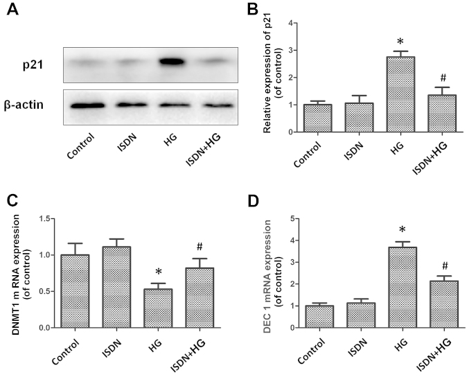
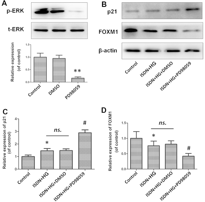
The present study was carried out to investigate the hypothesis that organic nitrates can attenuate the senescence of mesenchymal stem cells (MSCs), a superior cell source involved in the regeneration and repair of damaged tissue. MSCs were treated with high glucose (HG) in order to induce senescence, which was markedly attenuated by pre-treatment with isosorbide dinitrate (ISDN), a commonly used nitrate, as indicated by senescence-associated galactosidase (SA-β-gal) activity, p21 expression, as well as by the mRNA levels of DNA methyltransferase 1 (DNMT1) and differentiated embryo chondrocyte expressed gene 1 (DEC1), which are senescence-related biomarkers. It was also found that the senescent MSCs (induced by HG glucose) exhibited a marked downregulation in ERK activity and forkhead box M1 (FOXM1) expression, which was reversed by ISDN preconditioning. Of note, the inhibition of ERK phosphorylation or the downregulation of FOXM1 statistically abolished the favourable effects of ISDN. In addition, the investigation of the senescence-associated miR-130 family suggested that miR-130b mediates the beneficial effects of ISDN; it was found that the protective effects of ISDN against the senescence of MSCs were prominently reversed by the knockdown of miR-130b. Furthermore, the downregulation of ERK phosphorylation or FOXM1 expression decreased the miR-130b expression level; however, the suppression of miR-130b demonstrated no significant impact on ERK phosphorylation or FOXM1 expression. Taken together, to the best of our knowledge, the present study is the first to demonstrate the favourable effects of ISDN against HG-induced MSC senescence, which are mediated through the activation of the ERK/FOXM1 pathway and the upregulation of miR-130b.


Figures

Figure 1
Figure 1
High glucose (HG) accelerates mesenchymal stem cell (MSC) senescence in a concentration-dependent manner. MSCs were exposed to various concentrations of HG for 24 h or to mannitol (33 mM), which served as a negative control with an osmotic pressure equal to that of 33 mM HG. (A and B) Detection of p21 expression in MSCs by western blot analysis. The representative bands and statistical bar diagram are shown. The data are presented as the means ± standard error of the mean (SEM); n=5 in each experiment. (C and D) DNA methyltransferase 1 (DNMT1) mRNA expression and differentiated embryo chondrocyte expressed gene 1 (DEC1) mRNA expression were examined by quantitative PCR; the results are illustrated in the bar diagram. The data are presented as the means ± SE from 5 independent experiments. *P<0.05, **P<0.01 vs. the control group.
Figure 2
Figure 2
Isosorbide dinitrate (ISDN) does not have a notable effect on mesenchymal stem cell (MSCs) senescence. MSCs were cultured with ISDN at ISDN concentrations of 0 to 200 μM, and cellular senescence was compared between the groups. (A and B) p21 expression in MSCs was examined by western blot analysis; representative bands and a histogram are shown. The data are presented as the means ± standard error of the mean (SEM); n=5 in each group. (C and D) DNA methyltransferase 1 DNMT1 mRNA expression and differentiated embryo chondrocyte expressed gene 1 (DEC1) mRNA expression in MSCs was analyzed by quantitative PCR; the results are shown in the bar diagram. Data were obtained from 6 independent experiments, presented as the means ± SE, and compared with the control (untreated group; 0).
Figure 3
Figure 3
Isosorbide dinitrate (ISDN) preconditioning significantly attenuated the senescence of mesenchymal stem cells (MSCs) induced by high glucose (HG). MSCs were conditioned with or without ISDN (50 μM) for 6 h prior to HG treatment (33 mM) for an additional 24 h; cellular senescence was then investigated between the groups. (A and B) Cell aging was measured based on p21 expression, which was assessed by western blot analysis, and representative bands and a statistical histogram are shown. The results are presented as the means ± standard error of the mean (SEM); n=4 in each group. (C and D) Quantitative PCR was used to assess the mRNA expression of DNA methyltransferase 1 (DNMT1) and differentiated embryo chondrocyte expressed gene 1 (DEC1) in MSCs. The data are presented as the means ± SE and are shown in the histogram. Data were confirmed in 5 independent experiments. *P<0.05 vs. control; #P<0.05 vs. the HG group.
Figure 4
Figure 4
The ERK/FOXM1 pathway is affected by the induction of mesenchymal stem cell (MSC) senescence induced by high glucose (HG) and the protective effects of isosorbide dinitrate (ISDN). (A–D) MSCs were treated with HG at 15 mM or 33 mM or with mannitol (33 mM), which served as a negative control with an osmotic pressure equal to that of 33 mM HG. ERK phosphorylation and FOXM1 expression were evaluated by western blot, and representative bands are shown. The results are shown as the means ± standard error of the mean (SEM); n=3 per group. *P<0.05 vs. the control. (E–G) MSCs were preconditioned with ISDN (50 μM) for 6 h, followed by HG treatment (33 mM) for an additional 24 h. Western blot analysis was used to measure the activity of the ERK/FOXM1 pathway: representative bands and a statistical bar diagram are shown. The data are presented as the means ± SEM; n=3 in each experiment. *P<0.05 vs. the control group; #P<0.05 vs. the HG group.
Figure 5
Figure 5
Inhibition of ERK phosphorylation abolishes the favourable effects of isosorbide dinitrate (ISDN) on mesenchymal stem cell (MSC) senescence. (A) MSCs were treated with PD98059 (ERK inhibitor; 20 μM) or DMSO (served as negative control) for 30 min, and ERK phosphorylation was then investigated by western blot analysis. Representative bands and a corresponding bar diagram are shown. **P<0.01 vs. the control group. (B–D) MSCs were incubated with PD98059 (20 μM) or DMSO (served as negative control) for 30 min, followed by ISDN pre-culture for 6 h and high glucose (HG) treatment for an additional 24 h. Representative p21 and FOXM1 expression as determined by western blot analysis is shown, and the results are presented in the histogram. The data are presented as the means ± standard error of the mean (SEM); n=3 in each experiment. *P<0.05 vs. the control group; #P<0.05 vs. the ISDN + HG group; ns., not significant.
Figure 6
Figure 6
Knockdown of FOXM1 attenuates the beneficial effects of isosorbide dinitrate (ISDN) on mesenchymal stem cell (MSC) aging. (A) siRNA targeting FOXM1 or scrambled siRNA were transfected into the MSCs, and the representative bands and a histogram are shown with data presented as the means ± standard error of the mean (SEM) and confirmed in 3 independent experiments. **P<0.01 vs. control group. (B–D) After the knockdown of FOXM1, MSCs were cultured with ISDN for 6 h and then treated with high glucose (HG) for another 24 h. p21 expression and ERK phosphorylation levels were determined by western blot analysis and are statistically demonstrated in the histogram. Data are presented as the means ± SE from 3 independent experiments. *P<0.05 vs. the control group; #P<0.05 vs. the ISDN + HG group;.ns., not significant.
Figure 7
Figure 7
Isosorbide dinitrate (ISDN) pre-incubation delayed mesenchymal stem cell (MSC) senescence induced by high glucose (HG), which was mediated by the activation of the ERK/FOXM1 pathway. MSCs were incubated with PD98059 (ERK inhibitor; 20 μM) for 30 min prior to ISDN treatment (50 μM) for 6 h, followed by HG treatment (33 mM) for an additional 24 h. Alternatively, MSCs were treated with PD98059 or subjected to FOXM1 knockdown by siRNA and then treated with ISDN and HG. Senescence-associated β-galactosidase (SA-β-gal) activity and differentiated embryo chondrocyte expressed gene 1 (DEC1) and DNA methyltransferase 1 (DNMT1) mRNA expression were assessed to determine the cellular aging in each group. (A and B) Representative photomicrograph of the SA-β-gal assay is shown (scale bar, 20 μm). The percentage of β-galactosidase-positive cells (indicated by arrows) in each group was compared and is illustrated in the histogram; n=6 in each experiment. (C and D) Quantitative PCR analysis revealing the differences in DEC1 and DNA methyltransferase 1 (DNMT1) mRNA expression in each group. The data are presented as the means ± standard error of the mean (SEM); n=3 in each experiment. *P<0.05 vs. the control group; #P<0.05 vs. the HG group; &P<0.05 vs. the ISDN + HG group.
Figure 8
Figure 8
miR-130b, not miR-130a, is upregulated by the activation of the ERK/FOXM1 cascade during the protection of high glucose (HG)-induced mesenchymal stem cell (MSC) senescence by isosorbide dinitrate (ISDN). (A and B) MSCs were exposed to HG at progressively increasing concentrations for 24 h or to mannitol (33 mM), which served as a negative control with an osmotic pressure equal to that of 33 mM HG. The expression of the miR-130 family, including miR-130a and -130b, was determined by quantitative PCR and is shown in the histogram. The data are presented as the means ± standard error of the mean (SEM); n=3 in each experiment. *P<0.05, **P<0.01 vs. the control group. (C and D) MSCs were incubated with or without ISDN (50 μM) for 6 h, followed by HG treatment (33 mM) for an additional 24 h. The expression of miR-130a/b was analyzed by quantitative PCR, and the results are presented in the histogram. The data are presented as the means ± standard error of the mean (SEM); n=3 in each experiment. *P<0.05 vs. the control group; #P<0.05 vs. the HG group. (E and F) MSCs were cultured with PD98059 (20 μM) for 30 min prior to ISDN treatment (50 μM, 6 h), followed by HG treatment (33 mM, 24 h). Alternatively, MSCs were treated with PD98059 or subjected to FOXM1 knockdown by siRNA and then treated with ISDN and HG. miR-130b expression was evaluated by quantitative PCR, and the results are shown in the bar diagram. The data are presented as the means ± SEM; n=3 in each experiment. ns., not significant; &P<0.05 vs. the ISDN + HG group.
Figure 9
Figure 9
Downregulation of miR-130b abolishes the beneficial effects of isosorbide dinitrate (ISDN) on mesenchymal stem cell (MSC) aging. (A) The miR-130b level was quantified by quantitative PCR. The expression value of the control group was designated 1, and the levels of other groups were calibrated to this value. Data was presented as the means ± standard error of the mean (SEM) and confirmed in 5 independent experiments. **P<0.01 vs. control group. (B–D) Follwing the downregulation of miR-130b, MSCs were incubated with ISDN for 6 h and then exposed to high glucose (HG) for an additional 24 h. p21 expression was determined by western blot analysis. Quantitative PCR detection was further performed to reveal the differences in differentiated embryo chondrocyte expressed gene 1 (DEC1) and DNA methyltransferase 1 (DNMT1) mRNA expression in each group. The data are presented as the means ± SEM from 5 independent experiments. *P<0.05 vs. the ISDN + HG group; ns., not significant. (E–F) ERK phosphorylation and FOXM1 expression were determined following the suppression of miR-130b. Representative western blots are shown and the results are presented in the bar diagram as the means ± SEM from 5 independent experiments. NC, negative control.

Similar articles

Cited by

References

    1. Sung YH, Shin MS, Ko IG, et al. Ulinastatin suppresses lipopolysaccharide-induced prostaglandin E-2 synthesis and nitric oxide production through the downregulation of nuclear factor-κB in BV2 mouse microglial cells. Int J Mol Med. 2013;31:1030–1036. - PubMed
    1. Navascues J, Perez-Rontome C, Gay M, et al. Leghemoglobin green derivatives with nitrated hemes evidence production of highly reactive nitrogen species during aging of legume nodules. Proc Natl Acad Sci USA. 2012;109:2660–2665. doi: 10.1073/pnas.1116559109. - DOI - PMC - PubMed
    1. Matsui-Hirai H, Hayashi T, Yamamoto S, et al. Dose-dependent modulatory effects of insulin on glucose-induced endothelial senescence in vitro and in vivo: a relationship between telomeres and nitric oxide. J Pharmacol Exp Ther. 2011;337:591–599. doi: 10.1124/jpet.110.177584. - DOI - PubMed
    1. Ransohoff JD, Wu JC. Imaging stem cell therapy for the treatment of peripheral arterial disease. Curr Vasc Pharmacol. 2012;10:361–373. doi: 10.2174/157016112799959404. - DOI - PMC - PubMed
    1. Xu J, Qian J, Xie X, et al. High density lipoprotein cholesterol promotes the proliferation of bone-derived mesenchymal stem cells via binding scavenger receptor-B type I and activation of PI3K/Akt, MAPK/ERK1/2 pathways. Mol Cell Biochem. 2012;371:55–64. doi: 10.1007/s11010-012-1422-8. - DOI - PubMed

Publication types

MeSH terms