. 2014 Dec 19;289(51):35593-604.
doi: 10.1074/jbc.M114.620104. Epub 2014 Oct 29.

The E3 ubiquitin ligase thyroid hormone receptor-interacting protein 12 targets pancreas transcription factor 1a for proteasomal degradation

Affiliations

The E3 ubiquitin ligase thyroid hormone receptor-interacting protein 12 targets pancreas transcription factor 1a for proteasomal degradation

Naïma Hanoun et al. J Biol Chem. .

Abstract

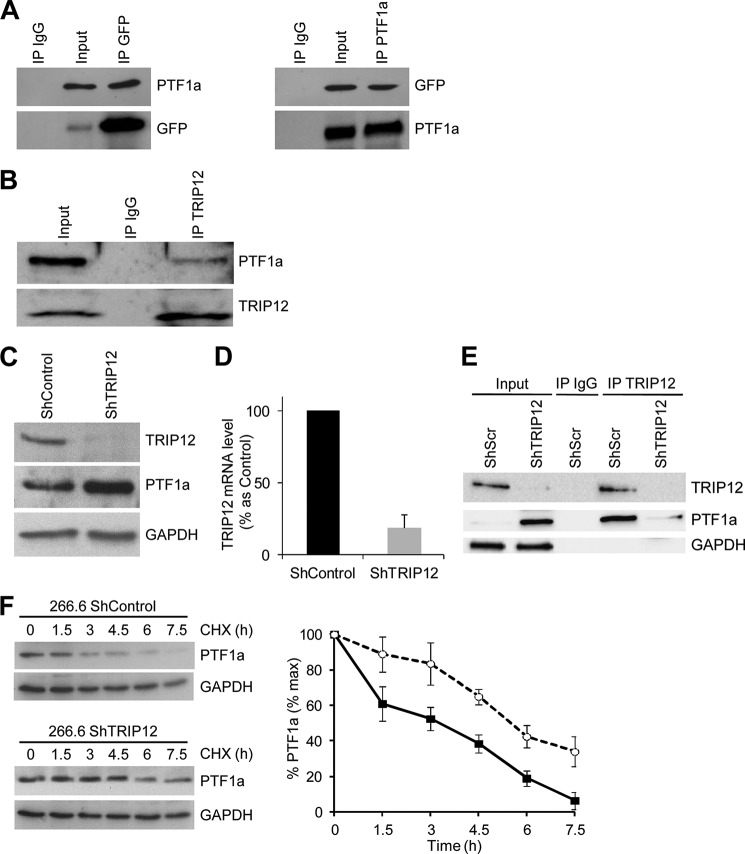
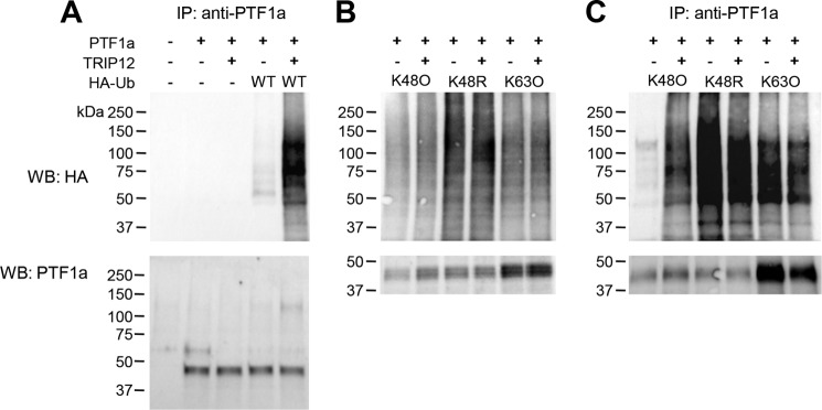
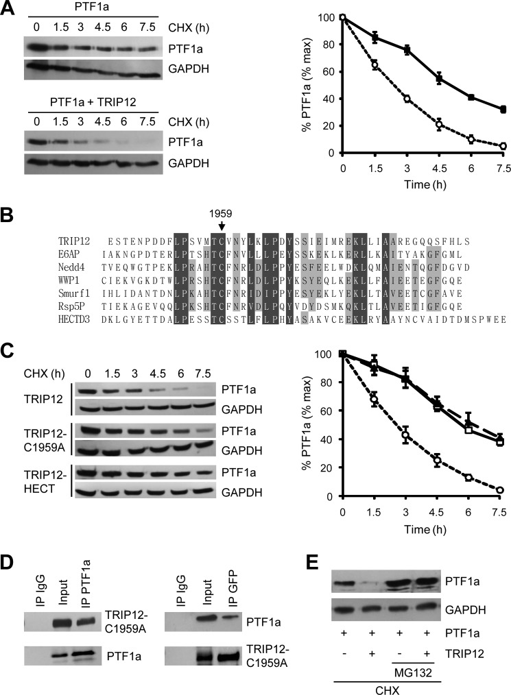
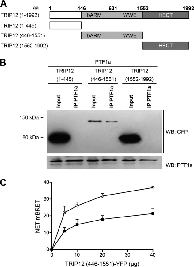
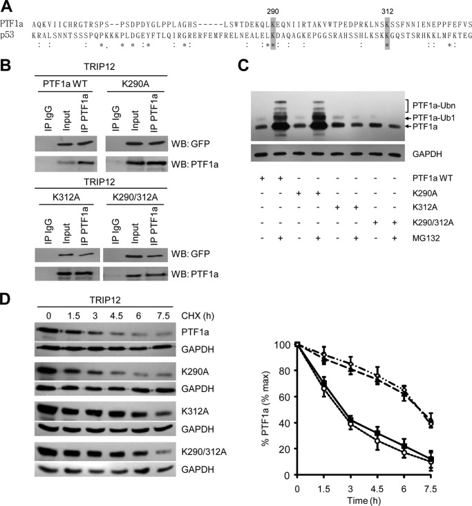
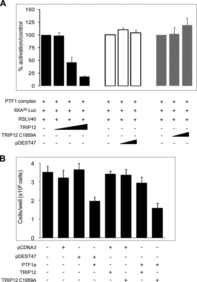
Pancreas transcription factor 1a (PTF1a) plays a crucial role in the early development of the pancreas and in the maintenance of the acinar cell phenotype. Several transcriptional mechanisms regulating expression of PTF1a have been identified. However, regulation of PTF1a protein stability and degradation is still unexplored. Here, we report that inhibition of proteasome leads to elevated levels of PTF1a and to the existence of polyubiquitinated forms of PTF1a. We used the Sos recruitment system, an alternative two-hybrid system method to detect protein-protein interactions in the cytoplasm and to map the interactome of PTF1a. We identified TRIP12 (thyroid hormone receptor-interacting protein 12), an E3 ubiquitin-protein ligase as a new partner of PTF1a. We confirmed PTF1a/TRIP12 interaction in acinar cell lines and in co-transfected HEK-293T cells. The protein stability of PTF1a is significantly increased upon decreased expression of TRIP12. It is reduced upon overexpression of TRIP12 but not a catalytically inactive TRIP12-C1959A mutant. We identified a region of TRIP12 required for interaction and identified lysine 312 of PTF1a as essential for proteasomal degradation. We also demonstrate that TRIP12 down-regulates PTF1a transcriptional and antiproliferative activities. Our data suggest that an increase in TRIP12 expression can play a part in PTF1a down-regulation and indicate that PTF1a/TRIP12 functional interaction may regulate pancreatic epithelial cell homeostasis.

Keywords: Basic Helix-Loop-Helix Transcription Factor (bHLH); E3 Ubiquitin Ligase; Pancreas; Pancreatic Cancer; Proteasome; Protein Degradation; Protein-Protein Interaction; Transcription Factor; Ubiquitylation (Ubiquitination); Yeast Two-Hybrid.


Figures

FIGURE 1.
FIGURE 1.
PTF1a is degraded by the ubiquitin-proteasome system. A, acinar AR4–2J cells expressing endogenous PTF1a and HEK-293T cells transfected with PTF1a vector were treated or not with MG132 (5 μm) for 10 and 16 h, respectively. Cell lysates were analyzed by Western blot using an anti-PTF1a antibody. B, acinar AR4–2J and 266.6 cells were treated with the protein synthesis inhibitor CHX (50 μg/ml) in the presence of MG132 (5 μm) for 4 h, and cell lysates were analyzed by Western blot using an anti-PTF1a antibody. C, acinar AR4–2J and 266.6 cells were treated with CHX in the presence or absence of the proteasome inhibitor lactacystin for 4 h, and cell lysates were analyzed by Western blot using anti-PTF1a antibody. D, HEK-293T cells were transfected with PTF1a expression vector and treated or not with MG132 for 10 h. Cell lysates were immunoprecipitated with anti-PTF1a antibody and immunoblotted with anti-PTF1a antibody (right panel) or anti-ubiquitin antibody (left panel); mono- and polyubiquitinated forms of PTF1a are indicated. Ub or Ubn, ubiquitin.
FIGURE 2.
FIGURE 2.
TRIP12 interacts with PTF1a and regulates PTF1a stability. A, co-immunoprecipitation of exogenous TRIP12 with exogenous PTF1a. HEK-293T cells were co-transfected with GFP-tagged TRIP12 and PTF1a expressing vectors. Cell lysates were immunoprecipitated or not (Input) with either nonspecific anti IgG, anti-GFP antibody, or anti-PTF1a antibody. The immunoprecipitates and lysates were analyzed by SDS-PAGE and subsequent immunoblotting with anti-PTF1a or anti-GFP antibodies. B, interaction between endogenous PTF1a and endogenous TRIP12. Acinar 266.6 cell lysates were immunoprecipitated or not (Input) with anti-TRIP12 antibody or with nonspecific IgG antibody and subsequently immunoblotted with anti-PTF1a or anti-TRIP12 antibodies C, TRIP12 and PTF1a protein levels in ShTRIP12 and ShControl 266.6 stable cell lines were measured by Western blot using appropriate antibodies. The level of GAPDH was used as loading control. D, TRIP12 and PTF1a mRNA level in ShTRIP12 and ShControl 266.6 stable cell lines was measured by quantitative RT-PCR using specific primers. The level of ribosomal protein S16 was used for normalization. The graph represents the means (± S.E.) of three different experiments. The results are expressed as percentages of levels in ShControl 266.6 cell line. E, TRIP12/PTF1a interaction in TRIP12 depleted 266.6 cells. Cell lysates from ShControl and ShTRIP12 stable 266.6 cell lines were immunoprecipitated or not (Input) with an anti-TRIP12 antibody. As control, cell lysate from ShControl 266.6 cell line was immunoprecipitated with a nonspecific IgG. Immunoprecipitated proteins were visualized by Western blot analysis using TRIP12 and PTF1a antibodies. The GAPDH level was used to verify equal amount of loaded protein in the Input lanes. F, PTF1a protein stability in TRIP12-depleted 266.6 cells. ShTRIP12 and ShControl 266.6 stable cell lines were treated with 10 μg/ml CHX for the indicated time. PTF1a protein level was analyzed by Western blot using an anti-PTF1a antibody. Relative amounts of PTF1a were calculated after normalizing to GAPDH protein level. Stability of PTF1a was determined from three separate experiments. Black squares represent ShControl 266.6 cell line, and white circles represent 266.6 ShTRIP12 cell line. IP, immunoprecipitation.
FIGURE 3.
FIGURE 3.
TRIP12 mediates Lys48-linked polyubiquitination of PTF1a. A, HEK-293T cells were co-transfected with PTF1a, WT HA-ubiquitin, and GFP-tagged TRIP12 expression plasmids as indicated. After MG132 exposure for 2 h, cell lysates were denatured and immunoprecipitated using anti-PTF1a antibodies, and equal amounts of PTF1a were immunoblotted with anti-HA antibody. B, HEK-293T cells were transfected with plasmids encoding HA-ubiquitin mutated at individual lysines except Lys48 (K48O), Lys63 (K63O), or mutated at Lys48 (K48R) as indicated. After MG132 exposure for 8 h, 50 μg of denatured cell lysates were loaded, HA-ubiquitinated proteins were detected with anti-HA antibody and PTF1a with anti-PTF1a antibody. C, HEK-293T cells were co-transfected with PTF1a, HA-ubiquitin mutants, and TRIP12 expression plasmids as indicated. After MG132 exposure for 8 h, cell lysates were denatured and immunoprecipitated using anti-PTF1a antibody. PTF1a was detected by immunoblotting with anti-HA or PTF1a antibodies. Standards molecular weights are indicated. IP, immunoprecipitation; WB, Western blot.
FIGURE 4.
FIGURE 4.
TRIP12 destabilizes and induces proteasome-dependent degradation of PTF1a. A, HEK-293T cells were treated with 50 μg/ml CHX for the indicated time after transfection with PTF1a and TRIP12-GFP. Fifty μg of cell lysates were analyzed by Western blot using an anti-PTF1a antibody, relative amounts of PTF1a were calculated after normalizing to GAPDH, and the half-life of PTF1a was determined from three separate experiments. Black squares, PTF1a; white circles, co-expression of PTF1a and TRIP12. B, sequence alignment of HECT domains from several E3 ubiquitin ligases, cysteine 1959 of TRIP12 is conserved. C, HEK-293T cells transfected with PTF1a and TRIP12 wild type, TRIP12-C1959A, or HECT domain expression vectors were analyzed by immunoblotting with anti-PTF1a antibody after treatment with CHX (50 μg/ml) as indicated. Relative amounts of PTF1a were calculated after normalizing to GAPDH, and the stability of PTF1a is represented from three separate experiments. White circles, co-expression of PTF1a and TRIP12 wild type; white squares, PTF1a and TRIP12-C1959A; black triangles, PTF1a and HECT domain. D, HEK-293T cells were co-transfected with GFP-tagged TRIP12-C1959A mutant and PTF1a expression vectors. Cell lysates were immunoprecipitated or not (Input) with anti-PTF1a antibody, anti-GFP antibody or nonspecific IgG antibody. The immunoprecipitates and cell lysates were analyzed by Western blot using anti-GFP or anti-PTF1a antibodies. E, HEK-293T cells were transfected with PTF1a expression vector in the presence or not of GFP-tagged TRIP12 construct and treated with CHX (50 μg/ml) in the presence or not of MG132 (5 μm) for 4 h, and cell lysates were analyzed by Western blot using an anti-PTF1a antibody. IP, immunoprecipitation.
FIGURE 5.
FIGURE 5.
The N-terminal region and the HECT domain of TRIP12 are not involved in PTF1a recognition. A, wild-type and truncated TRIP12 proteins. aa, amino acid. All constructs were GFP-tagged. B, HEK-293T cells were co-transfected with PTF1a and vectors for GFP-tagged truncated TRIP12 proteins as indicated. Cell lysates were immunoprecipitated or not (Input) with an anti-PTF1a antibody then analyzed by Western blot using anti-GFP or anti-PTF1a antibodies. A negative control consisting of anti-rabbit IgG beads alone led to no band (not shown). C, HEK-293T cells were transfected separately with the BRET donor (C- or N-terminally labeled PTF1a-HRluc vector) or the BRET acceptor (C-terminal labeled TRIP12 (446–1551) mutant-YFP vector). After 48 h, total cellular proteins were extracted. A total of 3 μg of lysate containing the BRET donor was mixed with variable amounts (0–40 μg) of lysate containing the BRET acceptor. White circles, C-terminal labeled PTF1a-HRluc vector; black squares, N-terminal labeled PTF1a-HRluc vector. The Rluc substrate coelenterazin-h was added 15 min before reading. The data represent the net BRET values expressed in NET milli BRET units. WB, Western blot.
FIGURE 6.
FIGURE 6.
Lysine 312 of PTF1a is required for ubiquitination and proteasome degradation. A, alignment of human PTF1a (amino acids 251–348) and p53 (amino acids 305–389) sequences. An asterisk indicates conserved amino acid residues, and a colon shows amino acid residues with similar properties. Gray boxes indicate lysine residues of PTF1a and p53 proteins located in similar amino acid environment. B, HEK-293T cells were co-transfected with GFP-tagged TRIP12 and PTF1a mutant expression vectors as indicated. Cell lysates were immunoprecipitated or not with an anti-PTF1a antibody or a nonspecific IgG antibody. The immunoprecipitates and lysates were analyzed by Western blot with anti-GFP or anti-PTF1a antibodies. C, HEK-293T cells transfected with wild-type PTF1a or PTF1a-K290A, PTF1a-K312A, and PTF1a-K290A/K312A mutants were treated or not with MG132 (5 μm) for 4 h. Cell lysates were analyzed by Western blot using an anti-PTF1a antibody, and mono- and polyubiquitinated forms of PTF1a are indicated. D, HEK-293T cells transfected with TRIP12 and the indicated PTF1a wild type and K290A, K312A, and K290A/K312A mutants were analyzed by immunoblotting with an anti-PTF1a antibody after treating with CHX (50 μg/ml) as indicated (left panel). Relative amounts of PTF1a were calculated after normalizing to GAPDH, and half-life of PTF1a was determined from three separate experiments in the right panel. White circles, co-expression of TRIP12 and PTF1a wild type; black squares, TRIP12 and PTF1a-K290A; black triangles, TRIP12 and PTF1a-K312A; white diamonds, TRIP12 and PTF1a-K290A/K312A.
FIGURE 7.
FIGURE 7.
TRIP12 reverses gene activation and antiproliferative activity of PTF1a. A, HEK-293T cells were transiently co-transfected with PTF1 complex (composed of PTF1a, RBPL, and HEB), the 6XA26-luc reporter construct that contains an hexamer of PTF1-responsive promoter and RSLV40 vector, in the presence of increasing amounts of TRIP12 (100, 200, and 500 ng/well) (black bars), TRIP12-C1959A (200 and 500 ng/well) (gray bars) or control pDEST47 plasmid (200 and 500 ng/well) (white bars). The RSLV40 vector was included as an internal control for normalization. The results are expressed as percentages of activation (means ± S.E., n = 3), and each experiment is performed in triplicate. No significant differences on the transcriptional activity are observed when TRIP12 or TRIP-C1959A constructs were transfected in the absence of PTF1a (data not shown). B, HEK-293T cells were co-transfected with PTF1a and with TRIP12 or TRIP12-C1959A or their corresponding controls plasmids (as indicated), stopped, and counted 6 days post-transfection. The results of experiments performed in triplicate are represented as counted cells per well and expressed in means ± S.E. (n = 3).

Similar articles

Cited by

References

    1. Hershko A., Ciechanover A. (1998) The ubiquitin system. Annu. Rev. Biochem. 67, 425–479 - PubMed
    1. Rotin D., Kumar S. (2009) Physiological functions of the HECT family of ubiquitin ligases. Nat. Rev. Mol. Cell Biol. 10, 398–409 - PubMed
    1. Lee J. W., Choi H. S., Gyuris J., Brent R., Moore D. D. (1995) Two classes of proteins dependent on either the presence or absence of thyroid hormone for interaction with the thyroid hormone receptor. Mol. Endocrinol. 9, 243–254 - PubMed
    1. Kajiro M., Tsuchiya M., Kawabe Y., Furumai R., Iwasaki N., Hayashi Y., Katano M., Nakajima Y., Goto N., Watanabe T., Murayama A., Oishi H., Ema M., Takahashi S., Kishimoto H., Yanagisawa J. (2011) The E3 ubiquitin ligase activity of Trip12 is essential for mouse embryogenesis. PLoS One 6, e25871. - PMC - PubMed
    1. Park Y., Yoon S. K., Yoon J. B. (2009) The HECT domain of TRIP12 ubiquitinates substrates of the ubiquitin fusion degradation pathway. J. Biol. Chem. 284, 1540–1549 - PubMed

Publication types

MeSH terms