. 2014 Dec 1;42(21):13082-95.
doi: 10.1093/nar/gku1029. Epub 2014 Oct 29.

Efficient processing of abasic sites by bacterial nonhomologous end-joining Ku proteins

Affiliations

Efficient processing of abasic sites by bacterial nonhomologous end-joining Ku proteins

Ana de Ory et al. Nucleic Acids Res. .

Abstract

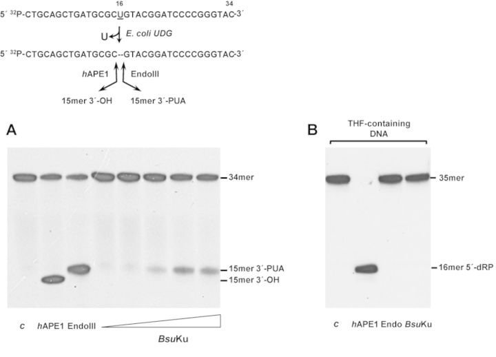
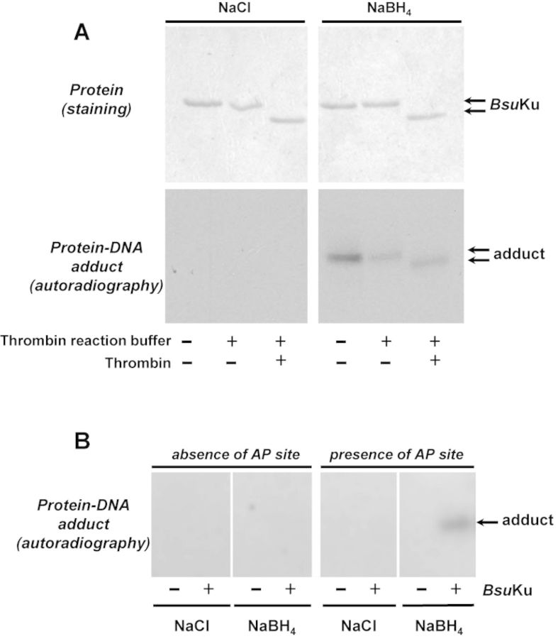
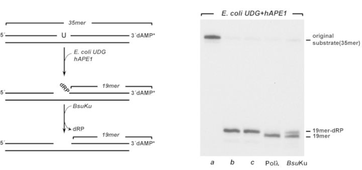
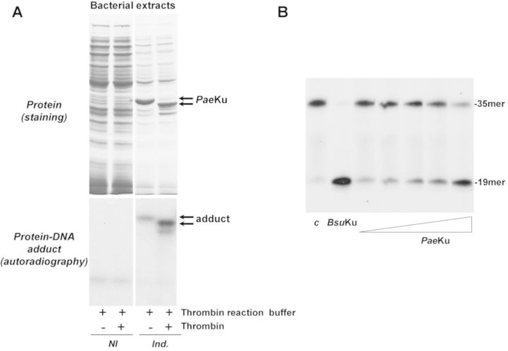
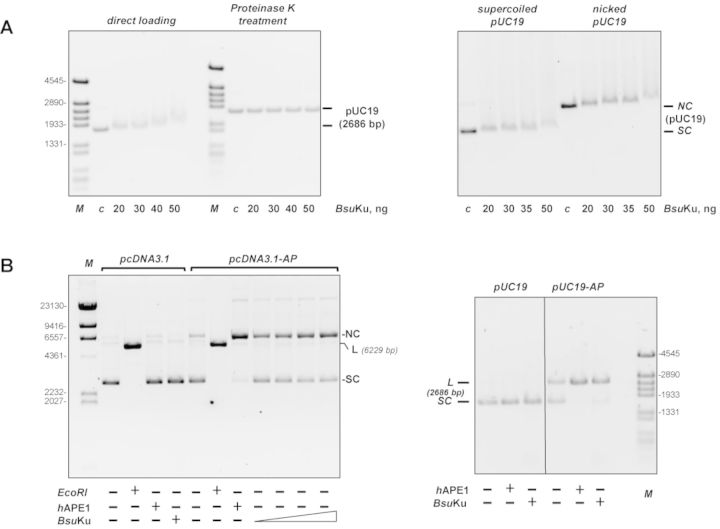
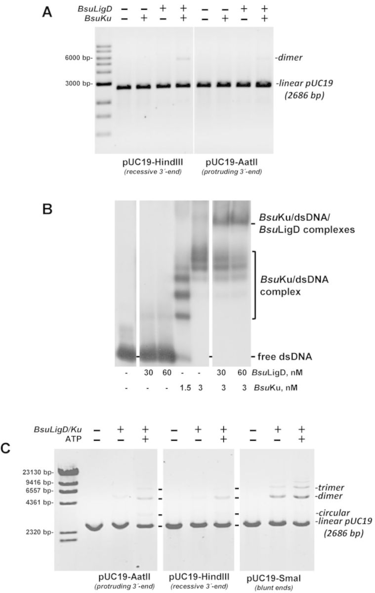
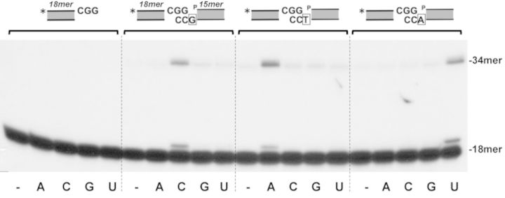
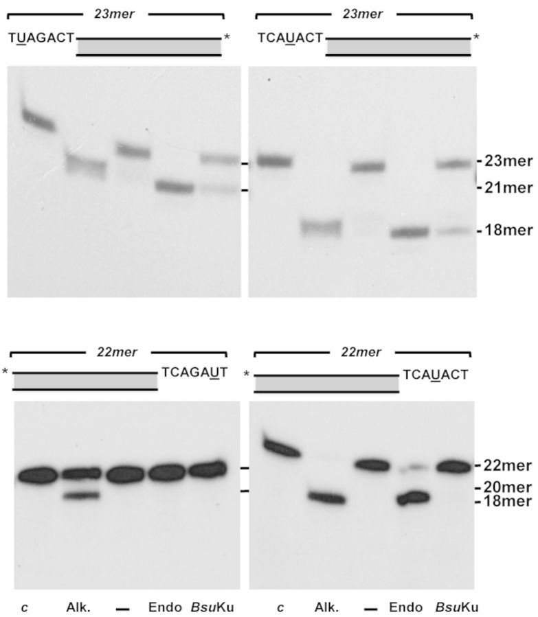
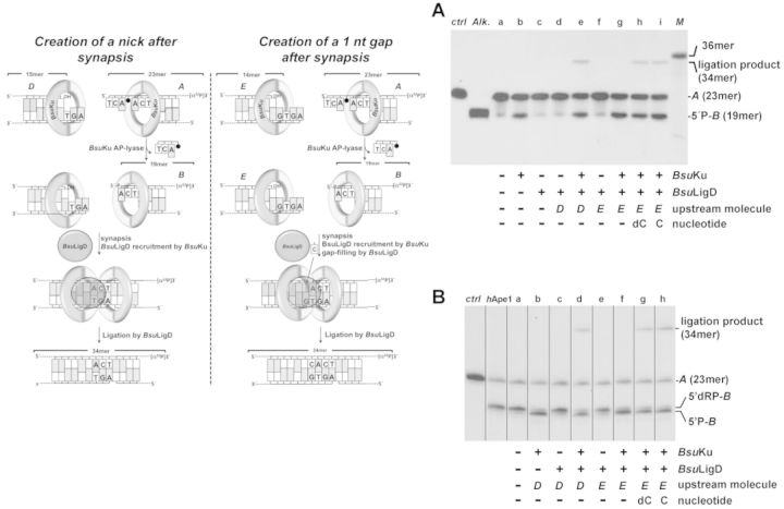
Intracellular reactive oxygen species as well as the exposure to harsh environmental conditions can cause, in the single chromosome of Bacillus subtilis spores, the formation of apurinic/apyrimidinic (AP) sites and strand breaks whose repair during outgrowth is crucial to guarantee cell viability. Whereas double-stranded breaks are mended by the nonhomologous end joining (NHEJ) system composed of an ATP-dependent DNA Ligase D (LigD) and the DNA-end-binding protein Ku, repair of AP sites would rely on an AP endonuclease or an AP-lyase, a polymerase and a ligase. Here we show that B. subtilis Ku (BsuKu), along with its pivotal role in allowing joining of two broken ends by B. subtilis LigD (BsuLigD), is endowed with an AP/deoxyribose 5'-phosphate (5'-dRP)-lyase activity that can act on ssDNA, nicked molecules and DNA molecules without ends, suggesting a potential role in BER during spore outgrowth. Coordination with BsuLigD makes possible the efficient joining of DNA ends with near terminal abasic sites. The role of this new enzymatic activity of Ku and its potential importance in the NHEJ pathway is discussed. The presence of an AP-lyase activity also in the homolog protein from the distantly related bacterium Pseudomonas aeruginosa allows us to expand our results to other bacterial Ku proteins.


Figures

Figure 1.
Figure 1.
BsuKu is endowed with AP-lyase activity. (A) The [32P]5′-labeled uracil-containing oligonucleotide was treated with Escherichia coli UDG, leaving an intact AP site. The resulting AP-containing DNA was incubated in the presence of either hAPE1 that cleaves 5′ to the AP site, EndoIII that incises 3′ to the AP site or increasing concentrations (9, 18, 36, 72 and 142 nM) of BsuKu for 1 h at 30°C, as described in Materials and Methods. c: control DNA after treatment with E. coli UDG. After incubation samples were analysed by 8 M urea-20% PAGE and autoradiography. Position of products is indicated. (B) The 3′[32P]3′-dAMP labeled oligonucleotide containing the lyase-resistant analog tetrahydrofuran (THF) at position 19 was incubated in the presence of either hAPE1, EndoIII or BsuKu (228 nM) as described above. c: control DNA incubated in the absence of proteins. Position corresponding to product (16mer 5′-dRP) is indicated.
Figure 2.
Figure 2.
Formation of BsuKu-DNA adducts. (A) Dependence of BsuKu-DNA cross-link on NaBH4. Reactions were performed as described in Materials and Methods, incubating 228 nM BsuKu with 3.1 nM of the 3′[32P]3′-dAMP labeled 35mer oligonucleotide containing an AP site at position 16 (after treatment with Escherichia coli UDG), in the presence of either 100 mM NaBH4 or NaCl (as indicated). Top panels, Coomassie blue staining after SDS-PAGE of purified BsuKu. Bottom panels, autoradiography of corresponding protein–DNA adducts after the SDS-PAGE separation shown in top panels. When indicated, protein was previously incubated with 0.05 U of thrombin at 20°C for 60 min. (B) Adduct formation is dependent on the presence of an abasic site. Reactions were performed as described in (A) but using as substrate 3.1 nM of the 3′[32P]3′-dAMP labeled 35mer oligonucleotide without removing the uracil at position 16 (absence of AP site) or after treatment with E. coli UDG (presence of AP site), in the presence of either 100 mM NaBH4 or NaCl (as indicated). Autoradiography of corresponding protein–DNA adduct after the SDS-PAGE separation is shown.
Figure 3.
Figure 3.
5′-dRP lyase activity of BsuKu. In vitro reconstitution of single-nucleotide BER. Left, schematic representation of a reconstituted BER reaction, indicating the different products formed. Right, autoradiogram illustrating BsuKu 5′-dRP lyase activity. a, original substrate; b, 5′-dRP-19mer product obtained after incubation with Escherichia coli UDG and hAPE1; c, 5′-dRP-19mer product incubated for 30 min at 30°C. Reactions were carried out as described in Materials and Methods in the presence of 228 nM BsuKu or Pol λ, as indicated.
Figure 4.
Figure 4.
AP-lyase activity is also present in the Ku protein from the Gram- bacteria P. aeruginosa. (A) Formation of PaeKu–DNA adducts. Top panels, expression of the recombinant PaeKu. For a better visualization of the induced PaeKu, 15 and 6 μg of total protein from the cellular extracts were loaded in the noninduced (NI) and in the induced (Ind.) lanes, respectively. Bottom panels, autoradiography of corresponding protein–DNA adducts after the SDS-PAGE separation. Reactions were performed as described in Materials and Methods, incubating either 1.7 μg of the noninduced bacterial extract or 0.7 μg of the induced extract with 3.1 nM of the 3′[32P]3′-dAMP labeled 35mer oligonucleotide containing an AP site at position 16 (after treatment with Escherichia coli UDG), in the presence of 100 mM NaBH4. When indicated, the bacterial extracts were previously incubated with 0.05 U of thrombin at 20°C for 60 min. (B) AP-lyase activity of purified PaeKu. 3.1 nM of the 3′[32P]3′-dAMP labeled 35mer oligonucleotide containing an AP site at position 16 (after treatment with E. coli UDG) was incubated in the presence of either BsuKu (228 nM), or increasing concentrations (14, 28, 57, 114 and 228 nM) of PaeKu for 1 h at 30°C, as described in Materials and Methods. c: control DNA after treatment with E. coli UDG. After incubation, samples were analysed by 8 M urea-20% PAGE and autoradiography. Position of products is indicated.
Figure 5.
Figure 5.
BsuKu binds circular DNA. (A) Left panel, BsuKu binding to covalently closed pUC19 plasmid. The assay was performed as described in Materials and Methods incubating the indicated amounts of BsuKu with 100 ng of supercoiled plasmid pUC19. Products were analysed on 0.7% agarose electrophoresis. One half of each reaction mixture was loaded directly onto the agarose gel (direct loading lanes), while the other half was being treated with Proteinase K. After proteolytic digestion, those samples were loaded in the same agarose gel (Proteinase K treatment lanes). The electrophoretical mobility of the pUC19 plasmid in each case is indicated. In lane c, DNA was incubated with 50 ng of BSA. Right panel, BsuKu binding to supercoiled and relaxed plasmid DNA. The assay was performed essentially as described above. (B) BsuKu AP lyase activity can act on circular substrates. The assay was performed as described in Materials and Methods, incubating either 142 nM of BsuKu with 100 ng of plasmid without AP sites (pcDNA3.1) or 18, 36, 72 and 142 nM of BsuKu with 100 ng of plasmid containing AP sites (pcDNA3.1-AP) (left panel). The absence and presence of the AP sites in pcDNA3.1 and pcDNA3.1-AP, respectively, was confirmed after digestion with hAPE1. As a control of the linear form, plasmids were also digested with EcoRI. NC: nicked circles; SC: supercoiled; L: linear. The assay shown in the right panel was performed by incubating 100 ng of plasmid without (pUC19) or with (pUC19-AP) AP sites with either hAPE1 or 142 nM of BsuKu, as indicated. Figure 5B, right panel is a composite image made from different parts of the same experiment.
Figure 6.
Figure 6.
(A) End joining of linearized plasmids. The assay was performed as described in Materials and Methods by incubating 100 ng of the specified digested pUC19 plasmid with 0.6 mM MnCl2 in the absence (-) or presence (+) of 142 nM of BsuLigD and BsuKu. After incubation for 60 min at 30°C, reactions were quenched by adjusting the mixtures to 0.2% SDS, 10 mM EDTA. Samples were incubated with 10 μg of proteinase K during 60 min at 37°C and further subjected to 0.7% agarose gel electrophoresis. The positions and size of DNA markers are indicated. (B) BsuKu recruits BsuLigD to DNA ends. Reactions were performed as described in Materials and Methods by incubating 1 nM of a 216 bp DNA fragment and the indicated concentrations of BsuLigD and BsuKu. For the supershift reactions, DNA was previously preincubated with BsuKu followed by the addition of the indicated amounts of BsuLigD. (C) Effect of ATP on the end-joining of linearized plasmids. The assay was performed essentially as described in (A). When indicated, 142 nM of BsuLigD/142 nM of BsuKu and/or 0.1 mM ATP were added.
Figure 7.
Figure 7.
End joining of partially complementary 3′-protruding DNA ends. The assays were carried out as described in Materials and Methods by incubating 1.25 nM of the labeled hybrid sp1CGG/sp1c depicted on top of Figure in the presence of 71 nM of both BsuLigD and BsuKu, 0.6 mM MnCl2, 2.5 nM of the nonlabeled hybrid (see Materials and Methods) indicated on top of Figure and 100 nM of the indicated NTP. The three DNA substrate molecules only differ in the marginal nucleotide indicated with open squares. After incubation for 30 min at 30°C, the elongation and ligation products were analysed by 8 M urea-20% PAGE and autoradiography. Asterisks indicate the 5′32P-labeled end of the primer strand.
Figure 8.
Figure 8.
BsuKu AP-lyase on protruding ends. The assay was performed as described in Materials and Methods. The substrates containing a uracil at the specified position within the 5′- (upper panels) or 3′- (lower panels) protruding ends were incubated with Escherichia coli UDG (c) to create an AP site in nearly all DNA molecules. After incubation of the AP-containing molecules with 2 units of E. coli Endo III (Endo), or 142 nM BsuKu for 30 min at 30°C, samples were analysed by 8 M urea-20% PAGE and autoradiography, as described in Materials and Methods. Positions corresponding to the products are indicated. Asterisks indicate either the 32P-5′ or the [32P]3′dAMP-3′end.
Figure 9.
Figure 9.
End joining of partially complementary DNA ends with near terminal AP sites. Left panel, schematic representation of the end-joining reaction, indicating the different substrates (filled circle represents an abasic site), products and the catalytic reactions that take place (see main text). (A) End joining of partially complementary DNA ends with near terminal AP sites; the assay was carried out as described in Materials and Methods by incubating 0.7 nM of the 3′-labeled substrate A and, when indicated 114 nM of BsuKu, 114 nM BsuLigD, 10 μM of the indicated nucleotide and 1 nM of the specified upstream substrate. After incubation for 30 min at 30°C, the products of the reaction were analysed by 8 M urea-20% PAGE and autoradiography (M: 36mer oligonucleotide used as size marker; ctrl: DNA after incubation with E. coli UDG; Alk. alkaline hydrolysis of the UDG-treated DNA). (B) End joining with a downstream DNA molecule bearing a protruding 5′-dRP end; the assay was performed as described in Materials and Methods by incubating 0.7 nM of the 3′-labeled substrate A with hAPE1. When indicated, the digested substrate was incubated with 114 nM of BsuKu, 114 nM of BsuLigD, 10 μM of the indicated nucleotide and 1 nM of the specified upstream substrate for 30 min at 30°C. After incubation, the products of the reaction were analysed by 8 M urea-20% PAGE and autoradiography. The lengths of the labeled substrate and the degradation and ligation products are indicated. Figure 9B is a composite image made from different parts of the same experiment.

Similar articles

Cited by

References

    1. Aguilera A., Gomez-Gonzalez B. Genome instability: a mechanistic view of its causes and consequences. Nat. Rev. Genet. 2008;9:204–217. - PubMed
    1. Daley J.M., Palmbos P.L., Wu D., Wilson T.E. Nonhomologous end joining in yeast. Annu. Rev. Genet. 2005;39:431–451. - PubMed
    1. Krejci L., Altmannova V., Spirek M., Zhao X. Homologous recombination and its regulation. Nucleic Acids Res. 2012;40:5795–5818. - PMC - PubMed
    1. Pitcher R.S., Brissett N.C., Doherty A.J. Nonhomologous end-joining in bacteria: a microbial perspective. Annu. Rev. Microbiol. 2007;61:259–282. - PubMed
    1. Shuman S., Glickman M.S. Bacterial DNA repair by non-homologous end joining. Nat. Rev. Microbiol. 2007;5:852–861. - PubMed

Publication types

MeSH terms