. 2014 Dec;3(12):1467-72.
doi: 10.5966/sctm.2014-0024. Epub 2014 Oct 29.

Efficient and cost-effective generation of mature neurons from human induced pluripotent stem cells

Affiliations

Efficient and cost-effective generation of mature neurons from human induced pluripotent stem cells

Cherif Badja et al. Stem Cells Transl Med. 2014 Dec.

Abstract

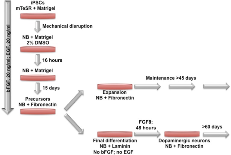
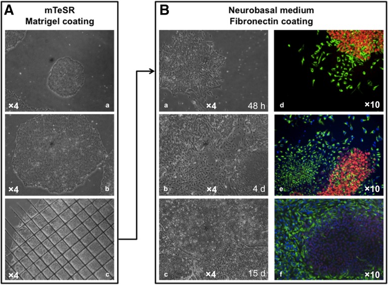
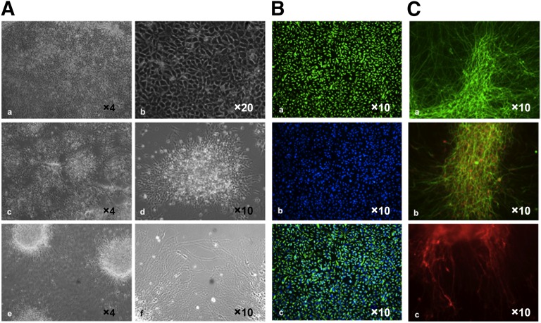
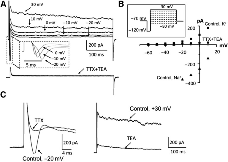
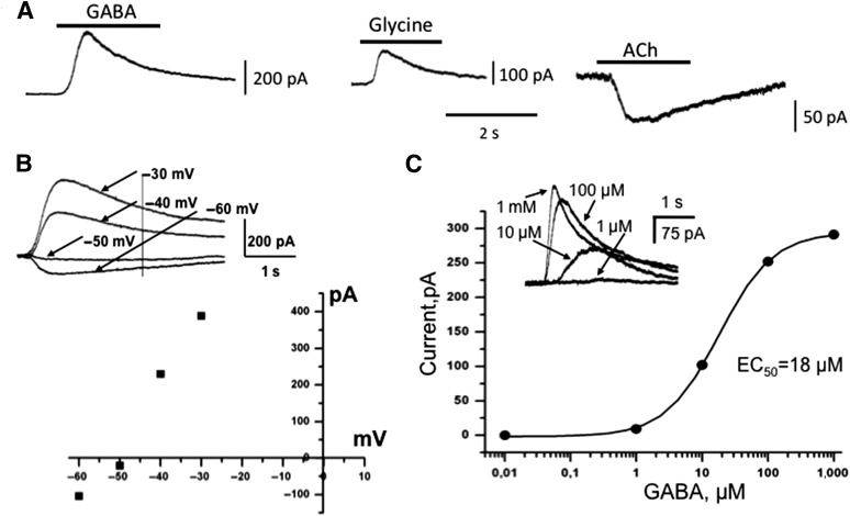
For years, our ability to study pathological changes in neurological diseases has been hampered by the lack of relevant models until the recent groundbreaking work from Yamanaka's group showing that it is feasible to generate induced pluripotent stem cells (iPSCs) from human somatic cells and to redirect the fate of these iPSCs into differentiated cells. In particular, much interest has focused on the ability to differentiate human iPSCs into neuronal progenitors and functional neurons for relevance to a large number of pathologies including mental retardation and behavioral or degenerative syndromes. Current differentiation protocols are time-consuming and generate limited amounts of cells, hindering use on a large scale. We describe a feeder-free method relying on the use of a chemically defined medium that overcomes the need for embryoid body formation and neuronal rosette isolation for neuronal precursors and terminally differentiated neuron production. Four days after induction, expression of markers of the neurectoderm lineage is detectable. Between 4 and 7 days, neuronal precursors can be expanded, frozen, and thawed without loss of proliferation and differentiation capacities or further differentiated. Terminal differentiation into the different subtypes of mature neurons found in the human brain were observed. At 6-35 days after induction, cells express typical voltage-gated and ionotrophic receptors for GABA, glycine, and acetylcholine. This specific and efficient single-step strategy in a chemically defined medium allows the production of mature neurons in 20-40 days with multiple applications, especially for modeling human pathologies.

Keywords: Dopaminergic neuron; GABA and glycine receptors; Human induced pluripotent cells; Neural differentiation; Neural induction; Neuronal progenitors; Patch clamp; Voltage-gated currents.


Figures

Figure 1.
Figure 1.
Schematic representation of the differentiation procedure. Days 1–15: differentiation of human iPSCs (hiPSCs) into human neuronal stem cells (hNSCs). The hiPSCs were expanded, and mature hiPSCs cultured in mTeSR on Matrigel-coated plates were mechanically disrupted in 30–50 small clumps using a 23-gauge needle and plated onto a Matrigel-coated 35-mm culture dish in differentiation medium (DM) supplemented with 20 ng/ml bFGF, 20 ng/ml EGF (DM+). Optimal results were obtained with 2% (v/v) DMSO for 16 hours. After overnight incubation, medium was replaced with DM+. Differentiated cells progressively emerge as a monolayer in the periphery of the hiPSC colony and can be maintained and expanded for up to 15 days with medium replacement every day. After 10–15 days of differentiation, cells at 90%–100% confluence are dissociated with Dispase. Small clumps of hNSCs were plated onto fibronectin-coated 35-mm culture dishes, and 90% of cells adhered within a few minutes after plating. hNSCs can be maintained for several passages or expanded after splitting with Accutase or a cell scraper and replating at a density of 1 × 10⁵ cells per 35-mm culture dish. For final differentiation, cells were mechanically separated with a 23-gauge needle and plated onto laminin-coated 6-well plates in DM without bFGF and EGF. Medium is replaced every day. Neurons develop in 5–7 days after plating. An example of final differentiation into dopaminergic neurons is presented after addition of specific cytokines, such as FGF8 and SHH. Abbreviations: bFGF, basic fibroblast growth factor; DMSO, dimethyl sulfoxide; EGF, epidermal growth factor; iPSC, induced pluripotent stem cell; NB, Neurobasal medium.
Figure 2.
Figure 2.
Differentiation of human induced pluripotent stem cells (hiPSCs) into neuronal progenitors. (A): Bright-field images of small immature (Aa) and mature (Ab) hiPSC colonies grown on Matrigel-coated plates in mTeSR before mechanical disruption (Ac). (B): After mechanical disruption using a 23-gauge needle, clumps of cells were plated on Matrigel and grown in differentiation medium. Left, bright-field images of neuronal differentiation at 48 hours (Ba), 4 days (Bb), or 15 days (Bc) after induction; (Bd) at 48 hours after induction, differentiated cells expressing the Nestin neuronal marker (green) migrate out of the OCT4-positive hiPSC colony (red); (Be) neuronal precursors express NESTIN (green) and hiPSCs express OCT4 at day 4 after induction; after 15 days (Bf), OCT4 expression is barely detectable, and Nestin-positive neuronal precursors reach 90%–100% confluence. Abbreviations: d, days; h, hours.
Figure 3.
Figure 3.
Production of mature neurons. (A): Illustration of the different steps of neuronal maturation. Neuronal progenitors can be expanded on solid-coated plates to 90%–100% confluence (magnification ×5 [Aa] and ×10 [Ab]) or dissociated and plated at a lower density (magnification ×5 [Ac] and ×10 [Ad]) for further differentiation (magnification ×5 [Ae] and ×10 [Af]). (B): Immunofluoresence staining 2 days after plating of neuronal progenitors on laminin in Neurobasal medium but without basic fibroblast growth factor and epidermal growth factor. The majority of cells express Nestin (Ba); cells were counterstained with 4′,6-diamidino-2-phenylindole (Bb) and merged (Bc). (C): At 20–30 days, mature neurons derived from hiPSCs express βIII-tubulin (green [Ca, Cb]) and the marker of mature neurons, NeuN (red [Cb]). Dopaminergic differentiation was induced by addition of SHH and FGF8, as described. The production and functionality of dopaminergic neurons were assessed by immunofluorescence staining with antibodies against tyrosine hydroxylase 15 days after induction (red [Cc]).
Figure 4.
Figure 4.
Electrophysiological properties of human induced pluripotent stem cell (hiPSC) differentiated neurons. (A): Representative example (upper panel) of currents evoked by depolarizing voltage steps indicated above the traces (see scheme of the protocol in 4B, insert); expanded from dashed lines (insert), rectangular traces of fast-activating, fast-inactivating inward currents evoked by depolarizing voltage steps, as indicated; example (bottom traces) of currents evoked by the same set of depolarizing voltage steps as indicated above after 1-minute preapplication of TTX (1 μM) plus TEA (20 mM). Note the strong inhibition of both inward and outward components. (B): Current-voltage relations of outward K+ (▪) and inward Na+ (▴) in control conditions and after 1-minute application of 1 μM TTX + 20 mM TEA (●); scheme of depolarizing protocol (insert) for recording current-voltage relations. In the different tests, pulse protocol was the same. (C): Examples of single traces illustrating separate inhibition of voltage-gated Na+ currents by 1 μM TTX (left) and K currents by 20 mM TEA (right). Note different time scales. Abbreviations: TEA, tetraethylammonium; TTX, tetrodotoxin.
Figure 5.
Figure 5.
Expression of neurotransmitter-activated channels in human induced pluripotent stem cell (hiPSC) differentiated neurons. (A): Representative examples of ionic currents induced by application of 1 mM GABA, 1 mM glycine, or 100 μM ACh in differentiated hiPSCs. Whole-cell recordings with intracellular solution containing 20 mM Cl at holding potential 0 mV (for GABA and glycine) and at −40 mV for ACh. Bars above traces indicate the time of agonist application. (B): Current-voltage relationship of the GABA-evoked inward current. Example (top) of superimposed traces of GABA-evoked currents at different potentials. Reversal potential was approximately −50 mV, as predicted from Nernst equation for intracellular solution containing 20 mM KCl. (C): Dose-response dependency and superimposed traces (insert) of whole-cell currents induced by rapid application of different concentration of GABA. Recording at 0 mV. Abbreviations: ACh, acetylcholine; EC50, half maximal effective concentration; s, seconds.

References

    1. Takahashi K, Yamanaka S. Induction of pluripotent stem cells from mouse embryonic and adult fibroblast cultures by defined factors. Cell. 2006;126:663–676. - PubMed
    1. Park IH, Zhao R, West JA, et al. Reprogramming of human somatic cells to pluripotency with defined factors. Nature. 2008;451:141–146. - PubMed
    1. Lee G, Papapetrou EP, Kim H, et al. Modelling pathogenesis and treatment of familial dysautonomia using patient-specific iPSCs. Nature. 2009;461:402–406. - PMC - PubMed
    1. Brennand KJ, Simone A, Jou J, et al. Modelling schizophrenia using human induced pluripotent stem cells. Nature. 2011;473:221–225. - PMC - PubMed
    1. Chamberlain SJ, Li XJ, Lalande M. Induced pluripotent stem (iPS) cells as in vitro models of human neurogenetic disorders. Neurogenetics. 2008;9:227–235. - PubMed

Publication types