. 2015 Jan;89(1):751-62.
doi: 10.1128/JVI.02430-14. Epub 2014 Oct 29.

Modulation of type I interferon-associated viral sensing during acute simian immunodeficiency virus infection in African green monkeys

Affiliations

Modulation of type I interferon-associated viral sensing during acute simian immunodeficiency virus infection in African green monkeys

Simon P Jochems et al. J Virol. 2015 Jan.

Abstract

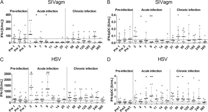
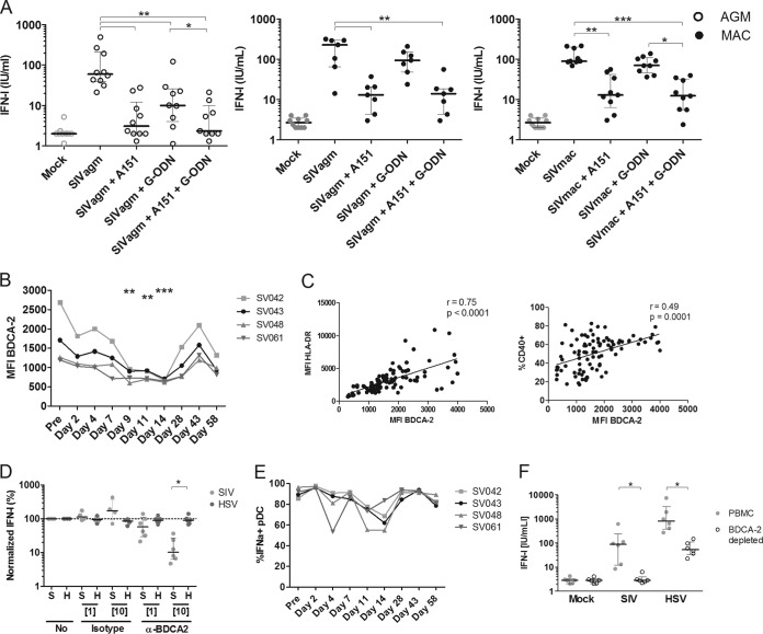
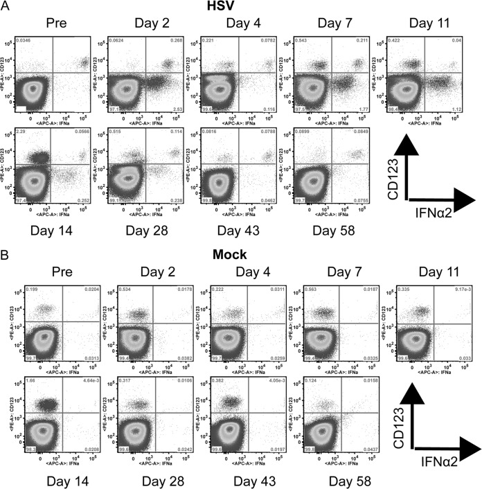
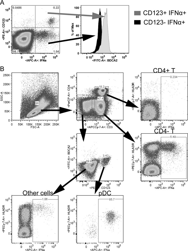
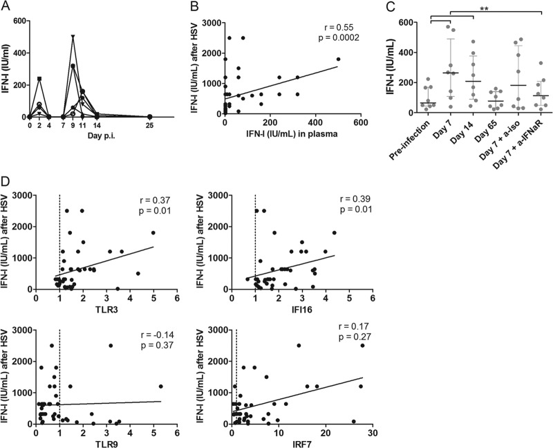
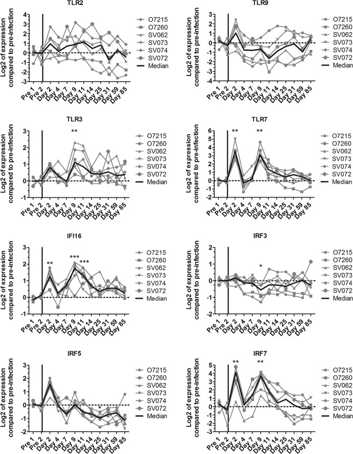
Natural hosts of simian immunodeficiency virus (SIV), such as African green monkeys (AGMs), do not progress to AIDS when infected with SIV. This is associated with an absence of a chronic type I interferon (IFN-I) signature. It is unclear how the IFN-I response is downmodulated in AGMs. We longitudinally assessed the capacity of AGM blood cells to produce IFN-I in response to SIV and herpes simplex virus (HSV) infection. Phenotypes and functions of plasmacytoid dendritic cells (pDCs) and other mononuclear blood cells were assessed by flow cytometry, and expression of viral sensors was measured by reverse transcription-PCR. pDCs displayed low BDCA-2, CD40, and HLA-DR expression levels during AGM acute SIV (SIVagm) infection. BDCA-2 was required for sensing of SIV, but not of HSV, by pDCs. In acute infection, AGM peripheral blood mononuclear cells (PBMCs) produced less IFN-I upon SIV stimulation. In the chronic phase, the production was normal, confirming that the lack of chronic inflammation is not due to a sensing defect of pDCs. In contrast to stimulation by SIV, more IFN-I was produced upon HSV stimulation of PBMCs isolated during acute infection, while the frequency of AGM pDCs producing IFN-I upon in vitro stimulation with HSV was diminished. Indeed, other cells started producing IFN-I. This increased viral sensing by non-pDCs was associated with an upregulation of Toll-like receptor 3 and IFN-γ-inducible protein 16 caused by IFN-I in acute SIVagm infection. Our results suggest that, as in pathogenic SIVmac infection, SIVagm infection mobilizes bone marrow precursor pDCs. Moreover, we show that SIV infection modifies the capacity of viral sensing in cells other than pDCs, which could drive IFN-I production in specific settings.

Importance: The effects of HIV/SIV infections on the capacity of plasmacytoid dendritic cells (pDCs) to produce IFN-I in vivo are still incompletely defined. As IFN-I can restrict viral replication, contribute to inflammation, and influence immune responses, alteration of this capacity could impact the viral reservoir size. We observed that even in nonpathogenic SIV infection, the frequency of pDCs capable of efficiently sensing SIV and producing IFN-I was reduced during acute infection. We discovered that, concomitantly, cells other than pDCs had increased abilities for viral sensing. Our results suggest that pDC-produced IFN-I upregulates viral sensors in bystander cells, the latter gaining the capacity to produce IFN-I. These results indicate that in certain settings, cells other than pDCs can drive IFN-I-associated inflammation in SIV infection. This has important implications for the understanding of persistent inflammation and the establishment of viral reservoirs.


Figures

FIG 1
FIG 1
Dynamics of IFN-I-producing capacity during SIVagm infection. We stimulated AGM PBMCs collected before and during the course of SIVagm infection and measured IFN-I production in the supernatant. The x axis displays the day postinfection at which cells were isolated and stimulated with SIV (A and B) or HSV (C and D). The y axis shows the total IFN-I produced after 18 h of stimulation (A and C) or the same IFN-I production corrected for the number of plasmacytoid dendritic cells among the PBMCs (B and D). The dashed horizontal line shows the median for all preinfection time point stimulations. The dashed vertical lines separate the stages of infection. Symbols represent individual animals (n = 5 to 11 for each time point). *, Wilcoxon, P < 0.05; **, Wilcoxon, P < 0.01. The P values indicate significant differences compared to the preinfection baseline.
FIG 2
FIG 2
Molecules involved in SIV sensing by AGM plasmacytoid dendritic cells. (A) PBMCs from 10 noninfected AGMs (open circles) and 7 to 9 MACs (closed circles) were stimulated with mock (gray) or SIVagm or SIVmac (black). A151 (5 μg/ml) and G-ODN (0.5 μM) were added at the same time as SIV to inhibit the TLR7 and TLR9 pathway, respectively. (B) Longitudinal follow-up of MFI levels of BDCA-2 on pDCs during early SIVagm infection. BDCA-2 expression was followed for four AGMs. (C) Expression levels of CD40 and HLA-DR were measured simultaneously on AGM pDCs. pDCs were defined as CD20 HLA-DR+ CD123+ BDCA2+ cells. r = Spearman's correlation. (D) anti-BDCA-2 or irrelevant IgG1 antibodies were added to PBMCs of six uninfected AGMs for 1 h at concentrations of 1 μg/ml and 10 μg/ml. After stimulation with SIVagm (S; light gray) or HSV-1 (H; dark gray) for 18 h, we measured IFN-I levels in the supernatants. IFN-I levels were normalized to IFN-I produced after no incubation with antibodies. The dotted line represents no difference compared to stimulation without antibodies. (E) Percentage of IFN-α2+ pDCs upon in vitro HSV stimulation at different time points of SIVagm infection. PBMCs were collected before and after SIVagm infection, and pDCs were analyzed for IFN-α2 production after in vitro stimulation. pDCs were defined as CD3 HLA-DR+ CD123+ cells. (F) BDCA-2-expressing cells were depleted to remove pDCs from PBMCs (n = 6 AGMs). Then, depleted (open circles) and whole (gray, filled circles) PBMCs were stimulated with SIV or HSV, after which IFN-I was measured in the supernatant. Symbols represent individual animals. *, Wilcoxon, P < 0.05; **, Wilcoxon, P < 0.01; ***, Wilcoxon, P < 0.001.
FIG 3
FIG 3
Non pDC-associated IFN-α production. Dot plots show CD123 and IFN-α2 expression on whole PBMCs collected throughout SIVagm infection. One representative AGM out of four analyzed is shown. (A) Cells after stimulation with HSV. (B) Mock-stimulated cells.
FIG 4
FIG 4
(A) Identification of cell types producing IFN-I upon HSV stimulation. (A) BDCA-2 expression on IFN-α2+ cells. PBMCs from a representative animal stimulated with HSV at day 2 p.i. are shown. From the CD123+ IFN-α+ (gray) and the CD123 IFN-α+ (black) quadrants, the expression of BDCA-2 is shown. (B) PBMCs were gated for distinct cell populations to identify cell types that produced IFN-α2. PBMCs from one AGM at day 2 p.i. are shown. A total of 5.5% of the CD4 T cells, 85.7% of the pDCs, and 1% of non-T, non-pDCs were IFN-α+ after HSV stimulation. All IFN-α-producing cells expressed high levels of HLA-DR. The identified cells producing IFN-α in this animal and at this time point are representative of data from days 2, 9, and 11 postinfection.
FIG 5
FIG 5
Auto-feedback loop of IFN-I during acute SIV infection of AGMs. (A) IFN-I levels present in the plasma of eight acutely SIV-infected AGMs. (B) Correlation between IFN-I levels in the plasma of AGMs during acute SIVagm infection in vivo and the amount of IFN-I produced upon in vitro HSV stimulation. (C) Plasma collected from healthy AGMs, acutely infected AGMs, and chronically infected AGMs was diluted to 5% in RPMI and added to PBMCs of eight healthy AGMs. After 4 h of incubation, cells were stimulated with HSV-1, and after another 18 h, supernatants were collected for IFN-I quantification. PBMCs were incubated with anti-IFN-αR2 or with an irrelevant antibody as a negative control at 10 μg/ml for 1 h to block IFN-α signaling before adding plasma. Symbols represent individual animals. (D) Correlation between mRNA levels of known HSV sensors in PBMCs of AGMs during acute SIVagm infection in vivo and the amount of IFN-I produced upon in vitro HSV stimulation of the same PBMCs. **, Wilcoxon, P < 0.01.
FIG 6
FIG 6
Gene expression profiles of genes involved in HSV sensing and IFN-I responses. mRNA levels were measured in PBMCs from six animals (the same animals as for Fig. 5) during SIVagm infection. The gray lines indicate individual animals, and the black lines show medians for all animals. *, Wilcoxon, P < 0.05; **, Wilcoxon, P < 0.01; ***, Wilcoxon, P < 0.001. The P values indicate a significant difference compared to the preinfection baseline.

Similar articles

Cited by

References

    1. Kirchhoff F. 2010. Immune evasion and counteraction of restriction factors by HIV-1 and other primate lentiviruses. Cell Host Microbe 8:55–67. doi:10.1016/j.chom.2010.06.004. - DOI - PubMed
    1. Fonteneau J-F, Larsson M, Beignon A-S, McKenna K, Dasilva I, Amara A, Liu Y-J, Lifson JD, Littman DR, Bhardwaj N. 2004. Human immunodeficiency virus type 1 activates plasmacytoid dendritic cells and concomitantly induces the bystander maturation of myeloid dendritic cells. J Virol 78:5223–5232. doi:10.1128/JVI.78.10.5223-5232.2004. - DOI - PMC - PubMed
    1. Romagnani C, Della Chiesa M, Kohler S, Moewes B, Radbruch A, Moretta L, Moretta A, Thiel A. 2005. Activation of human NK cells by plasmacytoid dendritic cells and its modulation by CD4+ T helper cells and CD4+ CD25hi T regulatory cells. Eur J Immunol 35:2452–2458. doi:10.1002/eji.200526069. - DOI - PubMed
    1. Karpov AV. 2001. Endogenous and exogenous interferons in HIV-infection. Eur J Med Res 6:507–524. - PubMed
    1. Asmuth DM, Murphy RL, Rosenkranz SL, Lertora JJ, Kottilil S, Cramer Y, Chan ES, Schooley RT, Rinaldo CR, Thielman N, Li XD, Wahl SM, Shore J, Janik J, Lempicki RA, Simpson Y, Pollard RB. 2010. Safety, tolerability, and mechanisms of antiretroviral activity of pegylated interferon Alfa-2a in HIV-1-monoinfected participants: a phase II clinical trial. J Infect Dis 201:1686–1696. doi:10.1086/652420. - DOI - PMC - PubMed

Publication types

MeSH terms

Substances