. 2015 Jan;89(1):676-87.
doi: 10.1128/JVI.02264-14. Epub 2014 Oct 29.

A newly isolated reovirus has the simplest genomic and structural organization of any reovirus

Affiliations

A newly isolated reovirus has the simplest genomic and structural organization of any reovirus

Albert J Auguste et al. J Virol. 2015 Jan.

Abstract

A total of 2,691 mosquitoes representing 17 species was collected from eight locations in southwest Cameroon and screened for pathogenic viruses. Ten isolates of a novel reovirus (genus Dinovernavirus) were detected by culturing mosquito pools on Aedes albopictus (C6/36) cell cultures. A virus that caused overt cytopathic effects was isolated, but it did not infect vertebrate cells or produce detectable disease in infant mice after intracerebral inoculation. The virus, tentatively designated Fako virus (FAKV), represents the first 9-segment, double-stranded RNA (dsRNA) virus to be isolated in nature. FAKV appears to have a broad mosquito host range, and its detection in male specimens suggests mosquito-to-mosquito transmission in nature. The structure of the T=1 FAKV virion, determined to subnanometer resolution by cryoelectron microscopy (cryo-EM), showed only four proteins per icosahedral asymmetric unit: a dimer of the major capsid protein, one turret protein, and one clamp protein. While all other turreted reoviruses of known structures have at least two copies of the clamp protein per asymmetric unit, FAKV's clamp protein bound at only one conformer of the major capsid protein. The FAKV capsid architecture and genome organization represent the most simplified reovirus described to date, and phylogenetic analysis suggests that it arose from a more complex ancestor by serial loss-of-function events.

Importance: We describe the detection, genetic, phenotypic, and structural characteristics of a novel Dinovernavirus species isolated from mosquitoes collected in Cameroon. The virus, tentatively designated Fako virus (FAKV), is related to both single-shelled and partially double-shelled viruses. The only other described virus in this genus was isolated from cultured mosquito cells. It was previously unclear whether the phenotypic characteristics of that virus were reflective of this genus in nature or were altered during serial passaging in the chronically infected cell line. FAKV is a naturally occurring single-shelled reovirus with a unique virion architecture that lacks several key structural elements thought to stabilize a single-shelled reovirus virion, suggesting what may be the minimal number of proteins needed to form a viable reovirus particle. FAKV evolved from more complex ancestors by losing a genome segment and several virion proteins.


Figures

FIG 1
FIG 1
Phase-contrast micrograph showing the cytopathic effects of CSW77 in representative cell lines: (A) negative control for C7/10; (B) C7/10 cells infected with CSW77. Note the significant syncytium formation and cell adhesion postinfection. (C) Typical plaques observed by CSW77 infection on C7/10 cells.
FIG 2
FIG 2
Neighbor-joining phylogeny of representative Reovirus sequences based on an amino acid alignment of the RdRp protein. Nodes are labeled with bootstrap values greater than or equal to 95%. FAKV is highlighted in bold. The scale bar indicates percent amino acid divergence. Members of the subfamily Spinareovirinae are highlighted in blue, and members of the subfamily Sedoreovirinae are highlighted in red. The various genera are also indicated in bold. Tip labels include the ICTV abbreviations. Sequences used for phylogenetic analyses are shown in Table S4 in the supplemental material.
FIG 3
FIG 3
Bayesian MCC tree for the subfamily Spinareovirinae based on 1,232 amino acids of the RdRp protein. Terminal branches of the tree are colored according to the trimer positions of the taxon at the tip. Internal branches are colored according to the most probable (modal) trimer positions of their parental nodes. All internal nodes had posterior probabilities (clade credibilities) of ≥0.95. Clamp proteins, their positions, and their probabilities of occurrence at the relevant nodes are indicated in bold. The number of genomic segments each genus contains is highlighted in color. The scale bar represents nucleotide substitutions per site.
FIG 4
FIG 4
The 7.8-Å structure of Fako virus determined by cryoelectron microscopy. (A) A typical micrograph collected with the DE-20 camera illustrates the preponderance of empty particles in preparations purified in TEN buffer. Particle boxes were extracted from each subframe, and particles were reconstituted by summing aligned, damage-corrected boxes. (B) The structure of the genome-free capsid was solved using the multipathway simulated annealing package and is colored radially. The inset shows a cutaway revealing the empty capsid interior and is labeled with radial measurements of the capsid outer surface, the turret peak, and the capsid inner surface. The structure of the FAKV full capsid was solved to 17 Å (C) to confirm that the absence of clamp near the 3-fold axis was not an artifact of genome release and agrees with the empty capsid structure (B). The inset shows a cutaway revealing ordered layers of dsRNA within the virion.
FIG 5
FIG 5
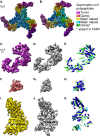
Protein composition of FAKV virions. Three segmented asymmetric units of FAKV (A) are colored by chain with different tints of the same color, representing symmetry-related copies of a protein. There are four proteins per asymmetric unit: a turret (purple) at the 5-fold axis, a dimer of the major capsid protein (blue and yellow), and a clamp protein (red) that binds to the major capsid protein surface. In contrast, the previously published structure of CPV (B) (11) and all other turreted reoviruses contain one clamp for each major capsid protein (red and green), and some have an additional clamp spanning the 2-fold axis. The turret protein (C to E), clamp protein (F to H), and major capsid protein (I to K) are contrasted between CPV and FAKV. The FAKV density map of each protein (C, F, I; colored as described above) was segmented from the 7.8-Å empty capsid reconstruction. A CPV map was obtained from the EM Data Bank (EMDB5256) (11) and filtered to similar resolution (D, G, J; gray). The FAKVα-helices (light green) and β-sheets (dark green) were compared to the α-helices (blue) and β-strands (cyan) of the CPV atomic structure (PDB 3IZX). While most secondary structural elements in the turret (E) and major capsid protein (K) are conserved, the clamp protein varies greatly around a conserved helix-barrel core (H).
FIG 6
FIG 6
Electrophoresis of FAKV proteins. (A) Purified FAKV virions were analyzed by SDS-PAGE and silver staining. Decreasing 10-fold dilutions in water of the same FAKV preparation are indicated by a triangle beneath lanes 1 to 3. (B) Putative open reading frames identified in the nine-segment genome of FAKV are colored to match their location in the virion. (C) Protein chains of one asymmetric unit of FAKV were segmented using Chimera and Gorgon. The folds and positions of these chains were similar to the turret, major capsid, or clamp of other Spinareovirinae. Open reading frames and protein chains are colored goldenrod and teal for two copies of the major capsid protein, purple for the turret protein, and red for the clamp protein. Lines indicate assignment of an SDS-PAGE band to a structural ORF.

References

    1. Weaver SC, Reisen WK. 2010. Present and future arboviral threats. Antiviral Res 85:328–345. doi:10.1016/j.antiviral.2009.10.008. - DOI - PMC - PubMed
    1. Attoui H, Mertens PPC, Becnel J, Belaganahalli S, Bergoin M, Brussard CP, Chappell Ciarlet JDM, del Vas M, Dermody TS, Dormitzer PR, Duncan R, Fcang Q, Graham R, Guglielmi KM, Harding RM, Hillman B, Makkay A, Marzachi C, Matthijnssens J, Milne RG, Mohd Jaafar F, Mori H, Noordeloos AA, Omura T, Patton JT, Rao S, Maan M, Stoltz D, Suzuki N, Upadhyaya NM, Wei C, Zhou H. 2012. Family Reoviridae, p 541–637. In King AMQ, Adams MJ, Carstens EB, Lefkowitz EJ (ed), Virus taxonomy: ninth report of the International Committee on Taxonomy of Viruses. Academic Press, London, United Kingdom.
    1. Miyazaki N, Uehara-Ichiki T, Xing L, Bergman L, Higashiura A, Nakagawa A, Omura T, Cheng RH. 2008. Structural evolution of Reoviridae revealed by Oryzavirus in acquiring the second capsid shell. J Virol 82:11344–11353. doi:10.1128/JVI.02375-07. - DOI - PMC - PubMed
    1. Reinisch KM, Nibert ML, Harrison SC. 2000. Structure of the reovirus core at 3.6 Å resolution. Nature 404:960–967. doi:10.1038/35010041. - DOI - PubMed
    1. Anzola JV, Xu ZK, Asamizu T, Nuss DL. 1987. Segment-specific inverted repeats found adjacent to conserved terminal sequences in wound tumor virus genome and defective interfering RNAs. Proc Natl Acad Sci U S A 84:8301–8305. doi:10.1073/pnas.84.23.8301. - DOI - PMC - PubMed

Publication types

MeSH terms

Associated data

LinkOut - more resources