. 2014 Feb;1(2):124-9.
doi: 10.1002/acn3.27. Epub 2014 Jan 9.

Hypertension drives parenchymal β-amyloid accumulation in the brain parenchyma

Affiliations

Hypertension drives parenchymal β-amyloid accumulation in the brain parenchyma

Celine Z Bueche et al. Ann Clin Transl Neurol. 2014 Feb.

Abstract

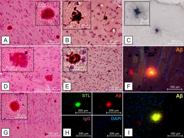
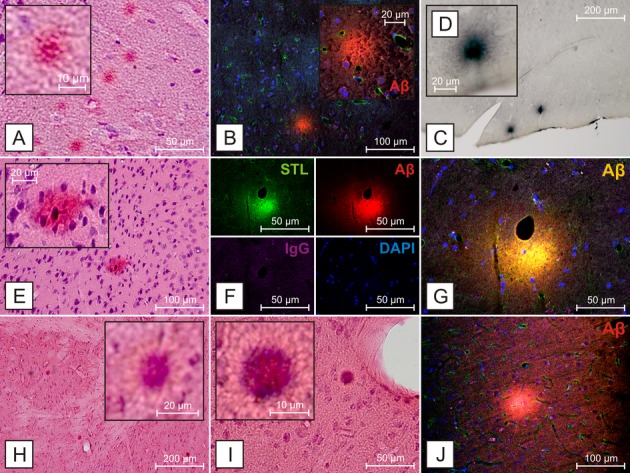
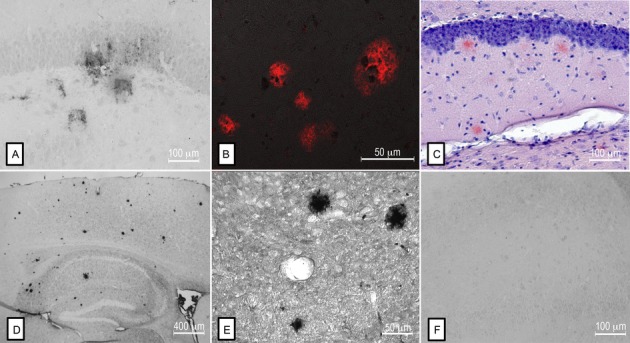
There is substantial controversy regarding the causative role of amyloid β (Aβ) deposition in Alzheimer's disease (AD). The cerebrovasculature plays an important role in the elimination of Aβ from the brain and hypertension is a well-known risk factor for AD. In spontaneously hypertensive stroke-prone rats (SHRSP), an animal model of chronic arterial hypertension, cerebral small vessel disease (CSVD) leads to age-dependent parenchymal Aβ accumulation similar to that observed in AD. These data approve the neuropathological link between CSVD and AD, confirm the challenge that parenchymal Aβ deposition is a specific marker for AD and disclose the meaning of SHRSP as valid experimental model to investigate the association between hypertension, CSVD, and Aβ plaques.


Figures

Figure 1
Figure 1
Aβ deposition in SHRSP 304, 26 weeks. Aβ deposition in cortical regions (A–C, E, G–I) and the basal ganglia (D, F) detectable using H&E (A, D, G) and Congo red (B, E) staining, as well as antibodies against Aβ (C) anti-human-Aβ clone 4G8, (F, H, I) anti-rodent-Aβ. Aβ-positive deposits do not colocalize with IgG (H) or DAPI (F, H, and I), suggesting that the staining is not due to leakage of the blood–brain barrier or nonspecific cellular labeling. Aβ deposits do colocalize with STL, as previously described (H). STL – Solanum tuberosum lectin, endothelial marker, DAPI – 4′,6-diamidino-2-phenylindole, for DNA detection.
Figure 2
Figure 2
Aβ deposition in SHRSP of different age groups. Hippocampal (A), cortical (B–D, E, J) and striatal (F–I) Aβ deposition in SHRSP of different age groups. Again, Aβ-positive deposits do not colocalize with IgG (F) or DAPI (B, F, G, and J), suggesting that the staining is not due to leakage of the blood–brain barrier or nonspecific cellular labeling. (A–D) SHRSP 331, 40 weeks, (E–G), SHRSP 309, 44 weeks, (H–J), SHRSP 308, 28 weeks, (A, E, H, I) H&E staining, (B, F, G, J) anti-rodent-Aβ, (C and D) anti-human-Aβ 1–17. STL – Solanum tuberosum lectin, endothelial marker, DAPI – 4′,6-diamidino-2-phenylindole, for DNA detection.
Figure 3
Figure 3
Comparison of Aβ deposition between SHRSP, Tg mouse models of AD, and human AD brains. Aβ-positive deposits in the hippocampus of a SHRSP (A) resemble those found in human AD brains (B). Similarly, plaques identified by Congo red staining (C) and by anti-Aβ immunohistochemistry (D, E) in mice overexpressing the Swedish human APP mutation, have a similar morphology to those identified in the SHRSP. Preabsorption of anti-Aβ (clone 4G8) with human Aβ40 attenuated the detection of Aβ staining in SHRSP (F).

References

    1. Gilbert BJ. The role of amyloid beta in the pathogenesis of Alzheimer's disease. J Clin Pathol. 2013;66:362–366. - PubMed
    1. Mawuenyega KG, Sigurdson W, Ovod V, et al. Decreased clearance of CNS beta-amyloid in Alzheimer's disease. Science. 2010;330:1774. - PMC - PubMed
    1. Bell RD, Zlokovic BV. Neurovascular mechanisms and blood-brain barrier disorder in Alzheimer's disease. Acta Neuropathol. 2009;118:103–113. - PMC - PubMed
    1. Carare RO, Hawkes CA, Jeffrey M, et al. Cerebral amyloid angiopathy, prion angiopathy, CADASIL and the spectrum of Protein Elimination-Failure Angiopathies (PEFA) in neurodegenerative disease with a focus on therapy. Neuropathol Appl Neurobiol. 2013;39:593–611. - PubMed
    1. Rodrigue KM, Rieck JR, Kennedy KM, et al. Risk factors for beta-amyloid deposition in healthy aging: vascular and genetic effects. JAMA Neurol. 2013;70:600–606. - PMC - PubMed

LinkOut - more resources