. 2014 Nov 25;53(46):7321-31.
doi: 10.1021/bi500916y. Epub 2014 Nov 13.

Biochemical, mechanistic, and spectroscopic characterization of metallo-β-lactamase VIM-2

Affiliations

Biochemical, mechanistic, and spectroscopic characterization of metallo-β-lactamase VIM-2

Mahesh Aitha et al. Biochemistry. .

Abstract

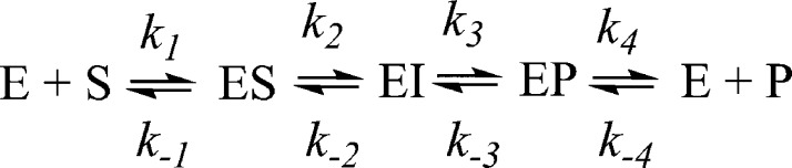
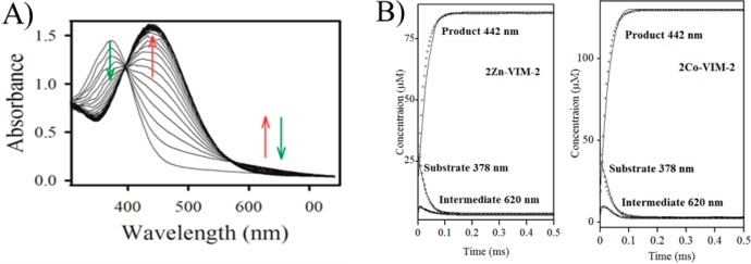
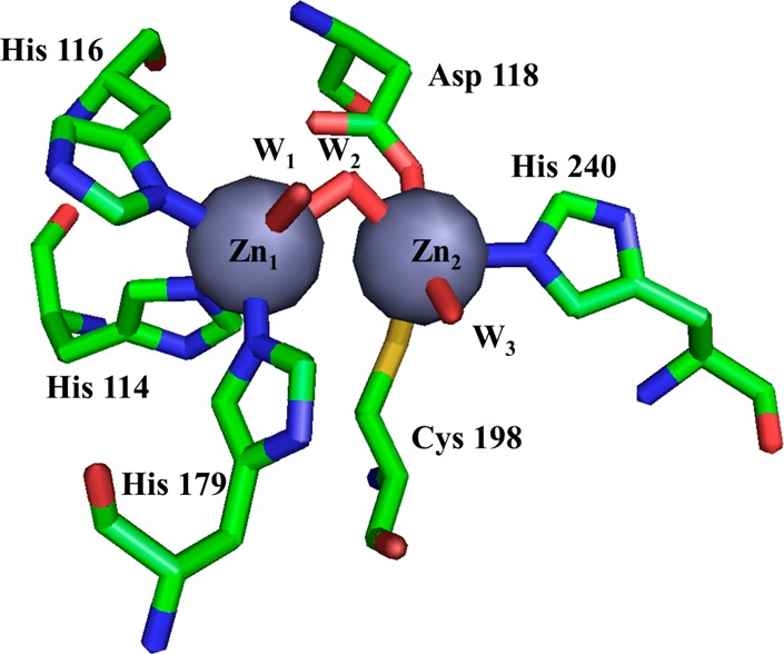
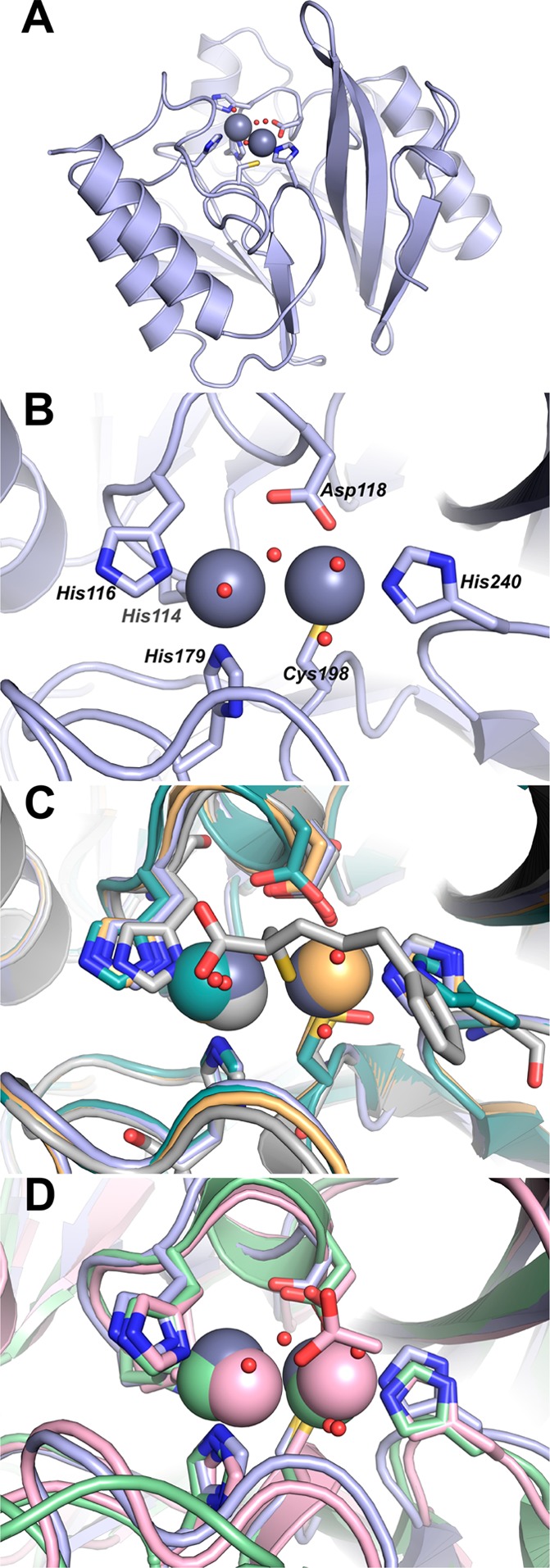
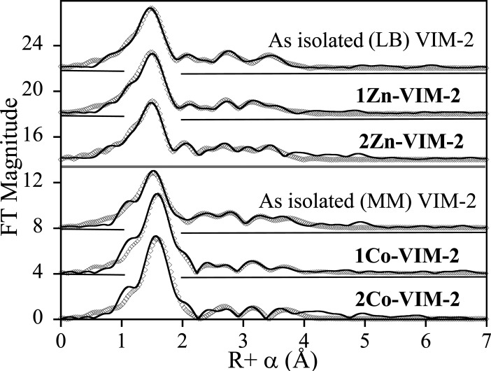
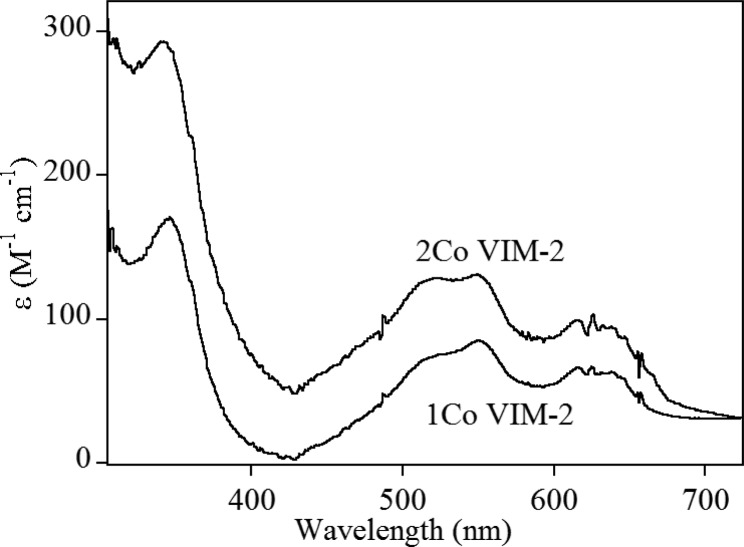
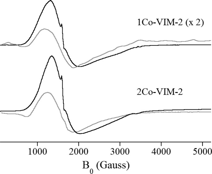
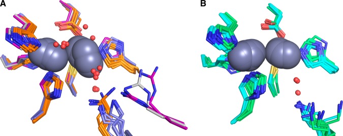
This study examines metal binding to metallo-β-lactamase VIM-2, demonstrating the first successful preparation of a Co(II)-substituted VIM-2 analogue. Spectroscopic studies of the half- and fully metal loaded enzymes show that both Zn(II) and Co(II) bind cooperatively, where the major species present, regardless of stoichiometry, are apo- and di-Zn (or di-Co) enzymes. We determined the di-Zn VIM-2 structure to a resolution of 1.55 Å, and this structure supports results from spectroscopic studies. Kinetics, both steady-state and pre-steady-state, show that VIM-2 utilizes a mechanism that proceeds through a very short-lived anionic intermediate when chromacef is used as the substrate. Comparison with other B1 enzymes shows that those that bind Zn(II) cooperatively are better poised to protonate the intermediate on its formation, compared to those that bind Zn(II) non-cooperatively, which uniformly build up substantial amounts of the intermediate.


Figures

Scheme 1
Scheme 1
Figure 1
Figure 1
(A) UV–vis spectra monitoring the reaction of 2Zn-VIM-2 (80 μM) and chromacef (80 μM) at pH 7.0 and 22 °C. (B) Progress curves for chromacef hydrolysis by 2Zn-VIM-2 (left) and 2Co-VIM-2 (right). Concentrations of substrate, product, and intermediate were calculated as described in Experimental Procedures. Theoretical progress curves are shown as open symbols, and the experimental progress curves are solid lines. The kinetic mechanism in Scheme 1 and the rate constants in Table 2 were used to generate the theoretical progress curves.
Figure 2
Figure 2
Active site from the VIM-2 crystal structure (PDB ID: 4NQ2) reported here. Zn1 is coordinated by three His and two water molecules, and Zn2 is coordinated by one His, one Cys, one Asp, and two water molecules. Zn1 and Zn2 are connected by a hydroxyl bridge.
Figure 3
Figure 3
Structure of 2Zn-VIM-2. (A) Cartoon representation of VIM-2 (pale blue). Active site side chains are shown as sticks; Zn(II) ions (gray) and water molecules (red) are shown as spheres. (B) Close-up highlighting coordination of the two Zn(II) ions by residues at the VIM-2 active site. A and B are from this study, PDB ID: 4NQ2. (C) Active site close-up for VIM-2 structures 4NQ2 (pale blue), 1KO2 (light teal), 1KO3 (light orange), and 2YZ3 (gray). Residues coordinating the Zn(II) ions including the 1KO2 cysteinesulfonic acid and the 2YZ3 mercaptocarboxylate inhibitor are shown as sticks; Zn(II) ions (pale blue, light teal, light orange, and gray) and water molecules are shown as spheres. (D) Active site close-up for VIM-2 (4NQ2, pale blue), IMP-1 (1DDK, light pink), and NDM-1 (3PSU, light green). Residues coordinating the Zn(II) ions and the Zn(II)-bound acetate ion from 1DDK are shown as sticks; Zn(II) ions (pale blue, light pink, and light green) and water molecules (red) are shown as spheres.
Figure 4
Figure 4
EXAFS Fourier-transforms for Zn(II)- and Co(II)-containing VIM-2 (solid lines) and corresponding best fits (open symbols). See Table 4, Tables S1–S6, and Figures S1–S6 for details. From top to bottom: recombinant LB VIM-2 (Zn K-edge), 1Zn-VIM-2 (Zn K-edge), 2Zn-VIM-2 (Zn K-edge), recombinant MM VIM-2 (Co K-edge), 1Co-VIM-2 (Co K-edge), and 2Co-VIM-2 (Co K-edge).
Figure 5
Figure 5
Optical spectra of 1Co- and 2Co-VIM-2.
Figure 6
Figure 6
X-band EPR spectra of 1Co- and 2Co-VIM-2, taken with B1B0 (gray lines) and B1B0 (black lines). The 1Co-VIM-2 spectra have been scaled by a factor of 2; all other sample and spectrometer conditions were identical.
Scheme 2
Scheme 2. Comparison of Nitrocefin (Left) and Chromacef (Right) Chromogenic Substrates
Figure 7
Figure 7
Comparison of available crystal structures for doubly zinc-loaded MBLs. (A) VIM-2 (PDB IDs: 4nq2 chain A, gray; 2yz3 chains A,B, purple; 1ko3 chain A, purple), IMP-1 (PDB IDs: 1dd6 chains A and B, orange; 1ddk chain A, orange; 1vgn chains A and B, orange; 2doo chains A and B, orange; 1jjt chains A and B, orange; and 1jje chains A and B, orange), and BcII (PDB IDs: 2uyx chain A, blue; 2bfx chains A and B, blue; 2bfl chains A and B, blue; and 2bg2 chains A and B, blue). (B) NDM-1 (PDB IDs: 4hl2 chains A and B, cyan; 4eyb chains A and B, cyan; 4exs chains A and B, cyan; 4exy chains A and B, cyan; 4ey2 chains A and B, cyan; 4eyf chains A and B, cyan; 4eyl chains A and B, cyan; 3spu chains B, C, D and E, cyan; and 3q6x chains A and B, cyan) and CcrA (PDB IDs: 1a7t chains A and B, green; 1a8t chains A and B, green; 2bmi chains A and B, green; and 1znb chains A and B, green). All zinc ions are represented as dark gray spheres. Structured waters within the active sites are shown as red spheres.

References

    1. CDC (2013) Antibiotic resistance threats in the United States, http://www.cdc.gov/drugresistance/threat-report-2013/pdf/ar-threats-2013.... - PubMed
    1. Nordmann P.; Naas T.; Poirel L. (2011) Global spread of carbapenemase-producing Enterobacteriaceae. Emerging Infect. Dis. 17, 1791–1798. - PMC - PubMed
    1. Watanabe M.; Iyobe S.; Inoue M.; Mitsuhashi S. (1991) Transferable imipenem resistance in Pseudomonas aeruginosa. Antimicrob. Agents Chemother. 35, 147–151. - PMC - PubMed
    1. Walsh T. R.; Toleman M. A.; Poirel L.; Nordmann P. (2005) Metallo-β-lactamases: the quiet before the storm?. Clin. Microbiol. Rev. 18, 306–325. - PMC - PubMed
    1. Bush K. (2013) The ABCD’s of β-lactamase nomenclature. J. Infect. Chemother. 19, 549–559. - PubMed

LinkOut - more resources