. 2015 Feb;23(2):235-43.
doi: 10.1038/mt.2014.214. Epub 2014 Oct 31.

Antiangiogenic variant of TSP-1 targets tumor cells in glioblastomas

Affiliations

Antiangiogenic variant of TSP-1 targets tumor cells in glioblastomas

Sung Hugh Choi et al. Mol Ther. 2015 Feb.

Expression of concern in

Abstract

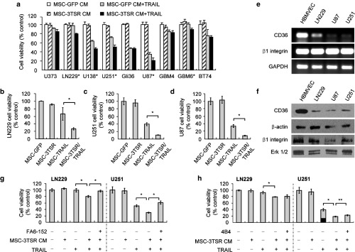
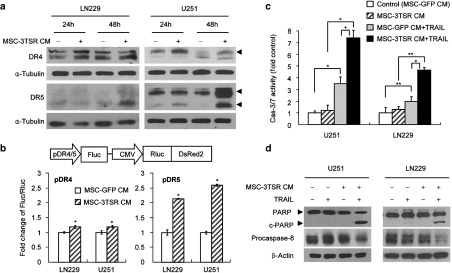
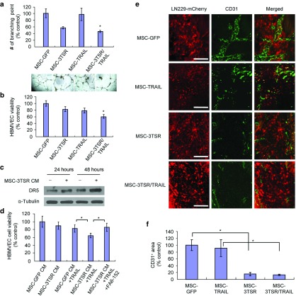
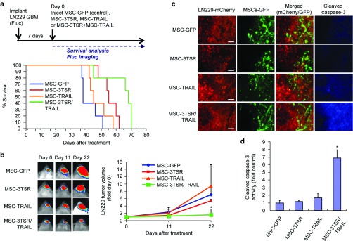
Three type-1 repeat (3TSR) domain of thrombospondin-1 is known to have anti-angiogenic effects by targeting tumor-associated endothelial cells, but its effect on tumor cells is unknown. This study explored the potential of 3TSR to target glioblastoma (GBM) cells in vitro and in vivo. We show that 3TSR upregulates death receptor (DR) 4/5 expression in a CD36-dependent manner and primes resistant GBMs to tumor necrosis factor-related apoptosis-inducing ligand (TRAIL)-induced caspase-8/3/7 mediated apoptosis. We engineered human mesenchymal stem cells (MSC) for on-site delivery of 3TSR and a potent and secretable variant of TRAIL (S-TRAIL) in an effort to simultaneously target tumor cells and associated endothelial cells and circumvent issues of systemic delivery of drugs across the blood-brain barrier. We show that MSC-3TSR/S-TRAIL inhibits tumor growth in an expanded spectrum of GBMs. In vivo, a single administration of MSC-3TSR/S-TRAIL significantly targets both tumor cells and vascular component of GBMs, inhibits tumor progression, and extends survival of mice bearing highly vascularized GBM. The ability of 3TSR/S-TRAIL to simultaneously act on tumor cells and tumor-associated endothelial cells offers a great potential to target a broad spectrum of cancers and translate 3TSR/TRAIL therapies into clinics.


Figures

Figure 1
Figure 1
MSC secreting 3TSR and TRAIL have synergistic cytotoxic effect on GBM cells in a CD36-dependent manner. (a) Cell viability was estimated using Celltiter Glow assay in different GBM lines. GBM lines were preincubated with MSC-3TSR-conditioned media for 24 hours and incubated with TRAIL (500 ng/ml for resistant lines, U373, LN229, U138, GBM4, GBM6, and BT74 and 100 ng/ml for sensitive lines, U251, Gli36, and U87) for additional 48 hours. *P < 0.01 between MSC-GFP CM + TRAIL and MSC-3TSR CM + TRAIL (Student's unpaired t-test). (b–d) Cell viability of LN229-mCherry-Fluc, U251-mCherry-Fluc, and U87-mCherry-Fluc was measured by bioluminescence imaging 120 hours (for LN229; TRAIL resistant), 72 hours (for U251; TRAIL semi-resistant), and 48 hours (for U87; TRAIL sensitive) after coculture with indicated MSCs in 1:4 ratio of MSCs to tumor cells. *P < 0.01 (e) Gene expression levels of CD36 and β1 integrin in GBM lines were measured by RT-PCR. (f) Western blotting showing the protein levels of glycosylated CD36 and β1 integrin. Glycosylated CD36 and β1 integrin were indicated by an arrowhead around 88 and 120 kD, respectively. ERK was used for loading control for β1 integrin. (g–h) LN229 and U251 GBM lines were preincubated with anti–CD36-blocking antibody FA6-152 or anti-β1 integrin blocking antibody 4B4 for 2 hours, cultured with MSC-GFP (control) or MSC-3TSR-conditioned media for 24 hours, and further incubated for 48 hours with TRAIL, 500 ng/ml for LN229 and 100 ng/ml for U251. Celltiter glow was used for cell viability assay. *P < 0.01 and **P < 0.05.
Figure 2
Figure 2
MSC-3TSR upregulates the expression of TRAIL receptors and primes TRAIL-resistant GBMs to caspase-8/3/7–mediated apoptosis. (a) LN229 and U251 were incubated with MSC-3TSR-conditioned media for indicated time, and DR4 and DR5 protein level was detected by western blotting. Arrowheads indicate DR4 (55 kD) and two different forms of DR5 (43 and 48 kD). (b) Schematic representation of polycistronic lentiviral vectors for dual bioluminescence reporter system to measure promoter activity of DR4 and DR5. Firefly luciferase (Fluc) and Renilla luciferase (Rluc) are expressed under the control of DR4 or DR5 promoter (pDR4/5) and CMV promoter, respectively. U251 and LN229 cells expressing the reporter system were incubated with MSC-3TSR-conditioned media for 48 hours. Transcriptional regulation of DR4 (bottom left) and DR5 (bottom right) by 3TSR were estimated by measuring bioluminescence of Fluc normalized with that of Rluc by Dual-Glo luciferase assay. *P < 0.01. (c) Caspase3/7 activation was assessed by caspase3/7 Glo assay in LN229 and U251 cells which were cultured with conditioned media from MSCs (GFP or 3TSR) for 48 hours and followed by treatment with TRAIL (50 ng/ml for U251 for 7 hours and 100 ng/ml for LN229 for 14 hours). *P < 0.01 and **P < 0.05 (Student's unpaired t-test). (d) Cleaved poly-ADP ribose polymerase and procaspase-8 protein level were western blotted to monitor apoptosis induction. Cell lysates were prepared from U251 and LN229 cells cultured in same condition as described in panel c.
Figure 3
Figure 3
MSC-3TSR inhibits angiogenesis and sensitizes brain endothelial cells to TRAIL in a CD36-dependent manner. (a) Photomicrographs and average branch points of HBMVEC after 18 hours coculture with U87 cells and MSCs expressing GFP (control), 3TSR, TRAIL, or 3TSR/TRAIL. *P < 0.01 between MSC-3TSR and MSC-3TSR/TRAIL. (b) Cell viability of HBMVEC was measured by Fluc bioluminescence after 48 hours post coculture with HBMVEC-Fluc-mCherry cells, U87 cells, and indicated MSCs. *P < 0.05 between MSC-TRAIL and MSC-3TSR/TRAIL. (c) Death receptor 5 expression was detected by western blot analysis using whole cell lysates of HBMVEC which was incubated with conditioned medium from MSC-3TSR or MSC-GFP (control) for indicated times. (d) HBMVEC was preincubated with anti-CD36 antibody FA6-152 for 1 hour before adding MSC-3TSR-conditioned media. Cell viability was measured by Celltiter Glo assay after 48 hours. *P < 0.05. (e) Immunofluorescence staining images of endothelial cell marker CD31 on brain sections from LN229-Fluc-mCherry GBM-bearing mice treated with indicated MSCs. MSCs were intratumorally injected 10 days after tumor implantation. Brain sections were prepared from mice sacrificed at day 5 after MSC injection. Bar = 100 µm. (f) Comparison of total area of tumor associated vessels obtained from anti-CD31 stained brain sections described in panel e (n = 3). *P < 0.01 (Student's unpaired t-test). The area of tumor associated vessels was measured using ImageJ.
Figure 4
Figure 4
MSC-3TSR/TRAIL inhibits growth of TRAIL-resistant GBM and prolongs survival of mice bearing TRAIL-resistant GBM. (a) Timeline and survival curves of mice bearing intracranial LN229-Fluc-mCherry GBM. MSC-GFP (control), MSC-3TSR, MSC-TRAIL, or MSC-3TSR/TRAIL were intratumorally injected 1 week after tumor implantation (n = 5 mice each group). Median survival is 38, 45, 55, and 68 days for MSC-GFP, MSC-TRAIL, MSC-3TSR, and MSC-3TSR/TRAIL, respectively. P < 0.05 between 3TSR/TRAIL group and other groups in log-rank test. (b) Representative bioluminescence images of tumor-bearing mice for each group used in panel a were shown (left). Change in tumor volumes was estimated by comparing Fluc bioluminescence intensity of mice at days 11 and 22 with that of day 0 (right). *P < 0.05 between MSC-3TSR/TRAIL and each individual group at day 22. (c) Immunofluorescence stained images of cleaved caspase-3 on brain sections from LN229-Fluc-mCherry GBM-bearing mice treated with indicated MSCs. MSCs were intratumorally injected 1 week after tumor implantation. Brain sections were prepared from mice sacrificed at 72 hours after MSC injection. Bar = 100 µm. (d) Comparison of immunofluorescence intensity of cleaved caspase-3 staining on brain sections imaged in panel c (n = 3). *P < 0.01 between MSC-3TSR/TRAIL and each individual group. Immunofluorescence intensity of cleaved caspase-3 from tumors was measured using ImageJ.

References

    1. Jain RK, di Tomaso E, Duda DG, Loeffler JS, Sorensen AG, Batchelor TT. Angiogenesis in brain tumours. Nat Rev Neurosci. 2007;8:610–622. - PubMed
    1. Plate KH, Mennel HD. Vascular morphology and angiogenesis in glial tumors. Exp Toxicol Pathol. 1995;47:89–94. - PubMed
    1. Rampling R, Cruickshank G, Lewis AD, Fitzsimmons SA, Workman P. Direct measurement of pO2 distribution and bioreductive enzymes in human malignant brain tumors. Int J Radiat Oncol Biol Phys. 1994;29:427–431. - PubMed
    1. Valk PE, Mathis CA, Prados MD, Gilbert JC, Budinger TF. Hypoxia in human gliomas: demonstration by PET with fluorine-18-fluoromisonidazole. J Nucl Med. 1992;33:2133–2137. - PubMed
    1. de Groot JF, Fuller G, Kumar AJ, Piao Y, Eterovic K, Ji Y.et al. (2010Tumor invasion after treatment of glioblastoma with bevacizumab: radiographic and pathologic correlation in humans and mice Neuro Oncol 12233–242. - PMC - PubMed

Publication types

MeSH terms

Substances

LinkOut - more resources