

Case Reports
doi: 10.1186/s13048-014-0101-7.
Borderline Brenner tumor of the ovary: a case report with immunohistochemical and molecular study
Affiliations
- PMID: 25358264
- PMCID: PMC4226905
- DOI: 10.1186/s13048-014-0101-7
Item in Clipboard
Case Reports
Borderline Brenner tumor of the ovary: a case report with immunohistochemical and molecular study
J Ovarian Res.
.
Abstract
Background: Borderline Brenner tumor of the ovary is a rare entity characterized by papillary structures with a fibro-vascular core, covered by a transitional epithelium, and by the absence of stromal infiltration. It is associated, by definition, with a benign component of Brenner tumor.
Case: We report a case of a 68-year-old woman, with a right ovarian mass, whose morphology and immuno-profile were consistent with the diagnosis of a borderline Brenner tumor. Immunohistochemistry carried out on selected markers may help to formulate the diagnosis, more than the molecular analyses.
Figures
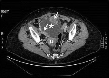
Contrast-enhanced CT scan showing a complex and heterogeneous pelvic mass with inner vegetations and septations in the anatomic site of the right adnexa (max diameter 17 cm), not dissociable from uterus and contiguous bowel (asterisk: pelvic mass; arrows: vegetations and septations; u: uterus).

Gross and microscopic description of Brenner tumor: a) Ovarian mass with solid papillary component; b) H & E representative of a solid adeno-fibromatous component (5X); c) H & E representative of cystic formations lined by mucinous columnar epithelium and by papillary transitional cellular component (10X); d) H & E representative of transitional epithelial cells with pale cytoplasm, indented nuclei and mild nuclear atypia (40X).
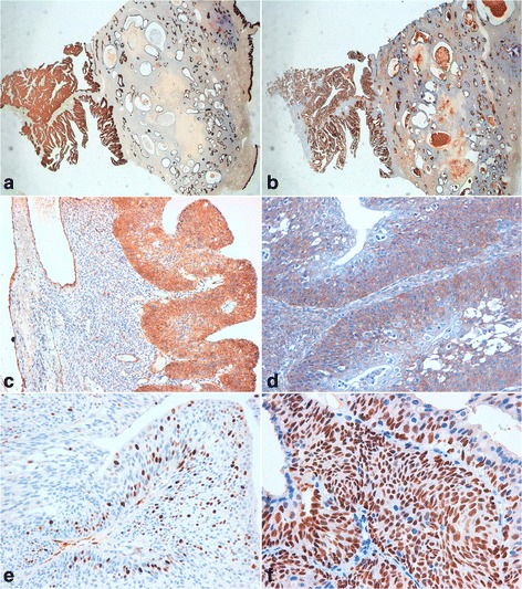
Borderline Brenner tumor immunoprofile. a) positive immunostaining for CK7 (10X); b) positive immunostaining for EMA (10X); c) positive immunostaining for thrombomodulin (20X); d) positive immunostaining for EGFR (20X); e) positive immunostaining for p16 (40X); f) positive immunostaining for p63 (20X).
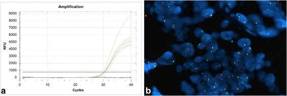
EGFR molecular analyses. a) Absence of mutations detected by RT-PCR method (the yellow increased curves indicate the internal controls of the kit, the blue flat lines indicate our sample); b) Absence of chromosomal aberrations detected by FISH method (red signals represent the EGFR gene and green signals represent the centromeric sequence in chromosome 7. The above picture shows a balanced disomy (each nucleus with 2 red and green signals, ratio = 1; Magnification, ×60)).
References
-
- Lee KR, Tavassoli FA, Prat J. Surface epithelial-stromal tumours. In: Tavassoli FA, Devilee P, editors. Pathology and Genetics: Tumours of the Breast and Female Genital Organs. World Health Organization Classification of Tumours 2003. Lyon: IARC Press; 2003. pp. 140–143.
-
- Cuatrecasas M, Catasus L, Palacios J, Prat J. Transitional cell tumors of the ovary: A comparative clinicopathologic, immunohistochemical, and molecular genetic analysis of Brenner tumors and transitional cell carcinomas. Am J Surg Pathol. 2009;33:556–67. doi: 10.1097/PAS.0b013e318188b84c. - DOI - PubMed
Publication types
MeSH terms
Substances
LinkOut - more resources
Full Text Sources
Other Literature Sources
Medical
Research Materials
Miscellaneous
