. 2014 Oct 30;55(11):7486-93.
doi: 10.1167/iovs.14-14792.

Acetylation preserves retinal ganglion cell structure and function in a chronic model of ocular hypertension

Affiliations

Acetylation preserves retinal ganglion cell structure and function in a chronic model of ocular hypertension

Oday Alsarraf et al. Invest Ophthalmol Vis Sci. .

Abstract

Purpose: The current studies investigate if the histone deacetylase (HDAC) inhibitor, valproic acid (VPA), can limit retinal ganglion cell (RGC) degeneration in an ocular-hypertensive rat model.

Methods: Intraocular pressure (IOP) was elevated unilaterally in Brown Norway rats by hypertonic saline injection. Rats received either vehicle or VPA (100 mg/kg) treatment for 28 days. Retinal ganglion cell function and number were assessed by pattern electroretinogram (pERG) and retrograde FluoroGold labeling. Western blotting and a fluorescence assay were used for determination of histone H3 acetylation and HDAC activity, respectively, at 3-day, 1-week, and 2-week time points.

Results: Hypertonic saline injections increased IOPs by 7 to 14 mm Hg. In vehicle-treated animals, ocular hypertension resulted in a 29.1% and 39.4% decrease in pERG amplitudes at 2 and 4 weeks, respectively, and a 42.9% decrease in mean RGC density at 4 weeks. In comparison, VPA treatment yielded significant amplitude preservation at 2 and 4 weeks and showed significant RGC density preservation at 4 weeks. No significant difference in RGC densities or IOPs was measured between control eyes of vehicle- and VPA-treated rats. In ocular-hypertensive eyes, class I HDAC activity was significantly elevated within 1 week (13.3 ± 2.2%) and histone H3 acetylation was significantly reduced within 2 weeks following the induction of ocular hypertension.

Conclusions: Increase in HDAC activity is a relatively early retinal event induced by elevated IOP, and suppressing HDAC activity can protect RGCs from ocular-hypertensive stress. Together these data provide a basis for developing HDAC inhibitors for the treatment of optic neuropathies.

Keywords: acetylation; neuroprotection; ocular hypertension; retina.


Figures

Figure 1
Figure 1
Schematic representation summarizing the key procedural aspects of the study. Each arrow corresponds to an important procedural time point, highlighting key experiments or measurements performed throughout the study. IOPs, intraocular pressure measurements; pERGs, pattern electroretinogram measurements; BID, twice daily; i.p., intraperitoneal; VPA, valproic acid.
Figure 2
Figure 2
Effect of valproic acid on IOP. (A) Baseline IOPs were measured 1 day prior to ocular hypertension induction (day −1). On day 0, ocular hypertension was induced (dashed line) by injecting approximately 50 μL 2.0 M hypertonic saline unilaterally in VPA-treated and vehicle-treated Brown Norway rats. Contralateral eyes served as controls. On days 3 through 28, significant (P < 0.001; n = 9) differences in IOPs were observed between ocular-hypertensive eyes and normotensive eyes in both vehicle and VPA treatment groups. No significant difference in IOPs was observed when comparing IOPs between vehicle- and VPA-treated groups in corresponding eyes. (B) Mean cumulative IOPs, calculated by addition of all IOP measurements with extrapolation for days unmeasured. The net result is the area under the curve of (A). No significant differences were noted between control and VPA-treated groups in hypertensive or normotensive eyes (n = 9). VPA, valproic acid; IOP, intraocular pressure.
Figure 3
Figure 3
Effect of ocular hypertension on retinal class I HDAC enzymatic activity. Extent of HDAC activity was examined by fluorescent detection of aminomethoxy-cumarin (AMC) following cleavage from enzymatically deacetylated lysines at 3 days, 1 week, and 2 weeks following ocular-hypertensive injury. Significant increases in HDAC activity were observed at 1 week (13.3 ± 2.2%) and 2 weeks (17.7 ± 1.9%) post ocular hypertension initiation. HDAC activity was presented as the percent activity change in hypertensive eyes relative to the contralateral control eyes. n = 4; *P < 0.05.
Figure 4
Figure 4
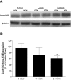
Effect of ocular hypertension on early changes in histone H3 acetylation. (A) Representative Western blot of retinal lysates for acetylated histone H3 normalized with β-actin. (B) Ratios of acetyl-histone H3 to β-actin were measured at 3 days, 1 week, and 2 weeks from the initiation of ocular hypertension. Ratio was expressed as a mean percentage of the hypertensive eyes relative to the control eyes (3 days, 95.5 ± 16.2%; 1 week, 74.2 ± 22.7%; 2 weeks, 61.9 ± 8.5%). n = 4; *P < 0.05. Acetyl-H3, acetyl-histone H3; HTN, hypertensive eye; NTN, normotensive eye.
Figure 5
Figure 5
Effect of HDAC inhibition on retinal histone H3 acetylation. Animals were treated with vehicle (n = 4) or valproic acid (VPA; 100 mg/kg, i.p.) (n = 4) 2 hours prior to tissue analysis, and a representative acetyl-histone H3 immunohistochemical stain is shown. Immunohistochemical staining for (A) retina acetylated histone H3; (B) nuclei staining (red) and (C) overlay of image of (A) and (B) in vehicle-treated animals; (D) retina acetylated histone H3; (E) nuclei staining (red) and (F) overlay of image of (D) and (E) in VPA-treated animals. i.p., intraperitoneal.
Figure 6
Figure 6
Effect of HDAC inhibition on functional neuroprotection using pattern electroretinography (pERG). Animals were treated with vehicle or valproic acid (VPA; 100 mg/kg; i.p.) twice daily on days 0 to 28, and resulting pERG amplitudes were recorded. (A) VPA administration showed significant preservation in raw pERG amplitudes compared to vehicle-treated counterparts at 2 weeks (8.9 ± 0.3 vs. 7.7 ± 0.5 μV) and 4 weeks (8.4 ± 1.0 vs. 6.1 ± 0.4 μV) post hypertension initiation. (B) Percentage reduction in pERG amplitudes of hypertensive eyes relative to normotensive contralateral control eyes (set at 0%). The decrease in pERG amplitudes of hypertensive eyes was calculated as a percentage of contralateral control eyes and adjusted by subtracting the absolute percentage change in amplitude of the contralateral control eyes from the respective baseline values. Vehicle-treated eyes demonstrated a 29.1 ± 1.9% and 39.4 ± 2.0% reduction at 2 weeks and 4 weeks, respectively. Eyes treated with VPA demonstrated significant preservation with 11.5 ± 1.7% and 12.4 ± 2.7% reduction at 2 weeks and 4 weeks, respectively (n = 10). (C) Representative pERG waveform from a control eye (blue) and ocular-hypertensive eye (black) at 4 weeks post injury initiation. Asterisk denotes significant difference between VPA-treated and vehicle-treated hypertensive groups. Data are expressed as mean ± SE. *P < 0.05; ψP < 0.01.
Figure 7
Figure 7
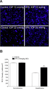
Effect of HDAC inhibition on RGC densities 5 days following injection of FluoroGold retrograde-labeling dye into the superior colliculi at day 28 of ocular hypertension. (A) Images of representative retinal flat mounts from all four groups with respective IOP measurements on day 28. (B) Vehicle-treated animals revealed a mean RGC density of 1951 ± 41 cells/mm2 for contralateral control eyes and a significant decrease to 1114 ± 36 cells/mm2 for hypertensive eyes. Animals treated with VPA demonstrated mean RGC densities of 1978 ± 38 and 1584 ± 60 cells/mm2 in normotensive and hypertensive eyes, respectively. Asterisks denote significant increases in labeled RGCs in hypertensive eyes from rats receiving VPA compared to animals receiving vehicle treatment (n = 10). *P < 0.05. RGC, retinal ganglion cell; VPA, valproic acid; BID, twice daily.

References

    1. Naskar R, Wissing M, Thanos S. Detection of early neuron degeneration and accompanying microglial responses in the retina of a rat model of glaucoma. Invest Ophthalmol Vis Sci. 2002; 43: 2962–2968. - PubMed
    1. Anderson DR. Normal-tension glaucoma (Low-tension glaucoma). Indian J Ophthalmol. 2011; 59 (suppl): S97–S101. - PMC - PubMed
    1. Quigley HA. Glaucoma. Lancet. 2011; 377: 1367–1377. - PubMed
    1. New M, Olzscha H, La Thangue NB. HDAC inhibitor-based therapies: can we interpret the code? Mol Oncol. 2012; 6: 637–656. - PMC - PubMed
    1. Biermann J, Grieshaber P, Goebel U, et al. Valproic acid-mediated neuroprotection and regeneration in injured retinal ganglion cells. Invest Ophthalmol Vis Sci. 2010; 51: 526–534. - PubMed

Publication types

MeSH terms

LinkOut - more resources