. 2014 Oct 14;3(4):634-49.
doi: 10.1016/j.stemcr.2014.07.010. Epub 2014 Sep 4.

Identification of a proximal progenitor population from murine fetal lungs with clonogenic and multilineage differentiation potential

Affiliations

Identification of a proximal progenitor population from murine fetal lungs with clonogenic and multilineage differentiation potential

Mélanie Bilodeau et al. Stem Cell Reports. .

Abstract

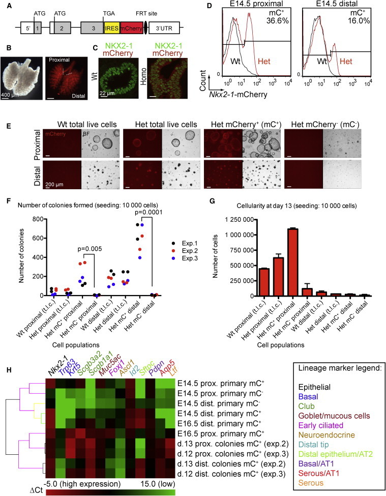
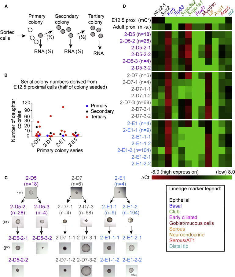
Lung development-associated diseases are major causes of morbidity and lethality in preterm infants and children. Access to the lung progenitor/stem cell populations controlling pulmonary development during embryogenesis and early postnatal years is essential to understand the molecular basis of such diseases. Using a Nkx2-1(mCherry) reporter mouse, we have identified and captured Nkx2-1-expressing lung progenitor cells from the proximal lung epithelium during fetal development. These cells formed clonal spheres in semisolid culture that could be maintained in vitro and demonstrated self-renewal and expansion capabilities over multiple passages. In-vitro-derived Nkx2-1-expressing clonal spheres differentiated into a polarized epithelium comprised of multiple cell lineages, including basal and secretory cells, that could repopulate decellularized lung scaffolds. Nkx2-1 expression thus defines a fetal lung epithelial progenitor cell population that can be used as a model system to study pulmonary development and associated pediatric diseases.


Figures

Figure 1
Figure 1
Colony-Formation Assay with E14.5 Proximal and Distal Lung Cells (A) mCherry knockin in the Nkx2-1 locus. Gray boxes indicate exons 1–3. UTR is shown in the open box. ATG or TGA indicates translation initiation or termination codon. (B) mCherry fluorescence detected by microscopy in the lungs of an E13.5 Nkx2-1mCherry mouse. (C) Cryosection immunostaining showing nuclear NKX2-1 correlating with cytoplasmic mCherry expression in the lung epithelium of an E13.5 homozygous (homo) Nkx2-1mCherry mouse. (D) Representative flow cytometry profiles of pulmonary cells harvested from WT or heterozygote (het) Nkx2-1mCherry mice. (E) Twelve-day-old colonies derived from sorted cell populations (p.1). BF, bright field. (F) Number of colonies observed for the indicated populations (scoring between days 6 and 12). Three independent experiments (Exp.) were performed in duplicate (dots). Significance was determined with a two-tailed Student’s t test (p value). t.l.c., total live cells. (G) Cellularity monitored at p.2 for a selected experiment. Values represent average of duplicate samples with SD. (H) Gene expression of selected markers monitored by quantitative real-time PCR in the indicated primary sorted cells or polyclonal colony populations. Values are normalized to the average of Actb and Hprt1 genes (ΔCt). ΔCt >15 may represent low or no expression. prox., proximal; dist., distal. See also Figures S1–S4 and Tables S2 and S3.
Figure 2
Figure 2
Colony Formation of E12.5 and E14.5 Primary Cells (A) Colony-formation assay with Nkx2-1-mC+ or Nkx2-1-mC cells sorted from proximal (Prox.) or distal (Dist.) lung regions at E12.5 or E14.5. The left panel shows the colony number from two experiments performed in duplicate (average and SD). The right panel shows the cellularity for experiment number 1 (average from two wells with SD). (B) Images of colonies at 17 days (p.1). (C) Immunostaining of mC+ colonies derived at E12.5 and E14.5 (16 days old, p.6). (D) Quantitative real-time PCR performed with RNA extracted from individual day 20 (p.2) mC+ colonies (col.) or primary sorted (mC+) or nonsorted (n.-s.) cells (n = 1 experiment). ΔCt heatmap with normalization of Ct values of indicated target genes to average of Actb and Hprt1 genes. The cutoff was set to a ΔCt of 10 (Ct 25–34 for reference genes). For the lineage marker legend, refer to Figure 1H. (E–H) Immunostaining of proximal lung epithelial cells from WT mice. Ctl+, positive control. See also Figure S5 for fractionation of proximal cells and Tables S2 and S3.
Figure 3
Figure 3
Clonogenicity and Differentiation of the Colonies Derived from E14.5 Proximal Lungs (A) Cell-mixing experiment performed with colonies derived from proximal lungs of E14.5 Nkx2-1mCherry or B5/EGFP mice (mCherry and EpCAM cell sorting, respectively). mC, mCherry. (B) Total colony number for each well (average and SD of duplicate wells; n = 1 experiment). (C) Representative colonies at day 12. (D) Scoring of colonies (8–12 days old) based on fluorescence at p.1 and p.2 in three independent wells. (E–I) Immunostaining with indicated antibodies of colonies derived from the proximal lung of E14.5 Nkx2-1mCherry mice (14–22 days old, p.2–p.6). See also Table S3.
Figure 4
Figure 4
In Vitro Self-Renewal of Primary Fetal Lung Cells (A) Serial passaging of colonies to assess in vitro self-renewal. (B) The capacity of isolated primary colonies to give rise to secondary colonies did not correlate with their sizes. Sixteen- to 20-day-old primary colonies were isolated at p.2–p.3 from two independent experiments (exp.) (sorting day = p.1). (C and D) Serial passaging of isolated primary and secondary colonies resulted in the formation of tertiary colonies through self-renewal expansion. For colonies derived from stage E12.5 (C) or E14.5 (D), primary colonies were isolated at p.2 or p.3 (16 days old) and secondary colonies at p.3 or p.4 (16 days old), respectively. Each dot represents the number of daughter colonies derived from a unique parental colony. For each clonal series, daughter colonies from four to five primary or from four to six secondary colonies were scored. (E) Paraffin section immunostaining of primary clones 3-G5 and 3-H5, and two daughter cultures for each (15–17 days old, p.6–p8). See also Table S3.
Figure 5
Figure 5
Self-Renewal Assessed with Individual Serial Colonies (A) In vitro self-renewal assay with serial single colonies and RNA extraction. (B) Number of secondary colonies derived from half of 18-day-old primary colonies (n = 4 primary colony series, p.1) and number of tertiary colonies derived from half of 23-day-old secondary colonies (n = 4–8 secondary colonies tested per series, p.2). (C) Images of colonies isolated in assay described in (B). Primary (1ary), secondary (2ary), and tertiary (3ary) colonies are 18 (p.1), 23 (p.2), and 22 days old (p.3), respectively. The number of daughter colonies is indicated in parentheses. (D) Quantitative real-time PCR with RNA isolated from colonies depicted in (C) from three serial series, as well as the initial sorted E12.5 mC+ proximal (prox.) cells and nonsorted (n.-s.) adult proximal tissue control. ΔCt heatmap is shown with normalization of Ct values of indicated target genes to Actb (ΔCt). The cutoff was set to a ΔCt of 8 (Ct 24.5–36.9 for Actb). See also Table S2.
Figure 6
Figure 6
Repopulation of Decellularized Lung Scaffolds with E14.5 Primary or Polyclonal Cultured Cells (A) Immunostaining of scaffolds repopulated with primary E14.5 Nkx2-1-mC+ cells freshly sorted from proximal lungs. (B) TEM showing secretory, ciliated, and basal cells in addition to epithelial polarization. Asterisk () indicates glycogen lakes. (C) Immunostaining (top) and electron microscopy (bottom) of scaffolds repopulated with polyclonal E14.5 mC+ cultured cells. The single asterisk () indicates that the matrix is representative of compact electron opaque and loosely associated filamentous material. The double asterisks (∗∗) indicate that the cytoplasm contains endoplasmic reticulum- and Golgi-derived membranes typical of a secretory cell. See also Figures S6 and S7 and Tables S1 and S3.
Figure 7
Figure 7
Repopulation of Decellularized Lung Scaffolds with Serial Clonal Cultures Derived from E14.5 Proximal Lungs (A) Section immunostaining of scaffolds repopulated with indicated Nkx2-1-mC+ clonal cultures. (B) SEM and TEM showing the epithelial morphology of clonal cultures in scaffolds. Overall, clonal cultures give rise to at least two to three distinct proximal cell types. Ø, decellularized lung matrix. See also Figures S6 and S7 and Tables S1 and S3.

References

    1. Alanis D.M., Chang D.R., Akiyama H., Krasnow M.A., Chen J. Two nested developmental waves demarcate a compartment boundary in the mouse lung. Nat. Commun. 2014;5:3923. - PMC - PubMed
    1. Broers J.L., de Leij L., Rot M.K., ter Haar A., Lane E.B., Leigh I.M., Wagenaar S.S., Vooijs G.P., Ramaekers F.C. Expression of intermediate filament proteins in fetal and adult human lung tissues. Differentiation. 1989;40:119–128. - PubMed
    1. Chang D.R., Martinez Alanis D., Miller R.K., Ji H., Akiyama H., McCrea P.D., Chen J. Lung epithelial branching program antagonizes alveolar differentiation. Proc. Natl. Acad. Sci. USA. 2013;110:18042–18051. - PMC - PubMed
    1. Daniely Y., Liao G., Dixon D., Linnoila R.I., Lori A., Randell S.H., Oren M., Jetten A.M. Critical role of p63 in the development of a normal esophageal and tracheobronchial epithelium. Am. J. Physiol. Cell Physiol. 2004;287:C171–C181. - PubMed
    1. Delplanque A., Coraux C., Tirouvanziam R., Khazaal I., Puchelle E., Ambros P., Gaillard D., Péault B. Epithelial stem cell-mediated development of the human respiratory mucosa in SCID mice. J. Cell Sci. 2000;113:767–778. - PubMed

Publication types