Review
. 2015 Mar;72(6):1029-43.
doi: 10.1007/s00018-014-1768-z. Epub 2014 Oct 31.

Mechanisms controlling neuromuscular junction stability

Affiliations
Review

Mechanisms controlling neuromuscular junction stability

Evelyne Bloch-Gallego. Cell Mol Life Sci. 2015 Mar.

Abstract

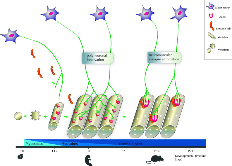
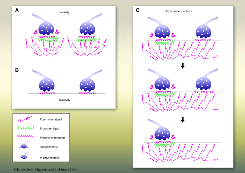
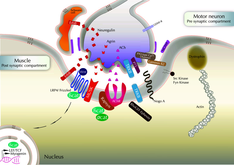
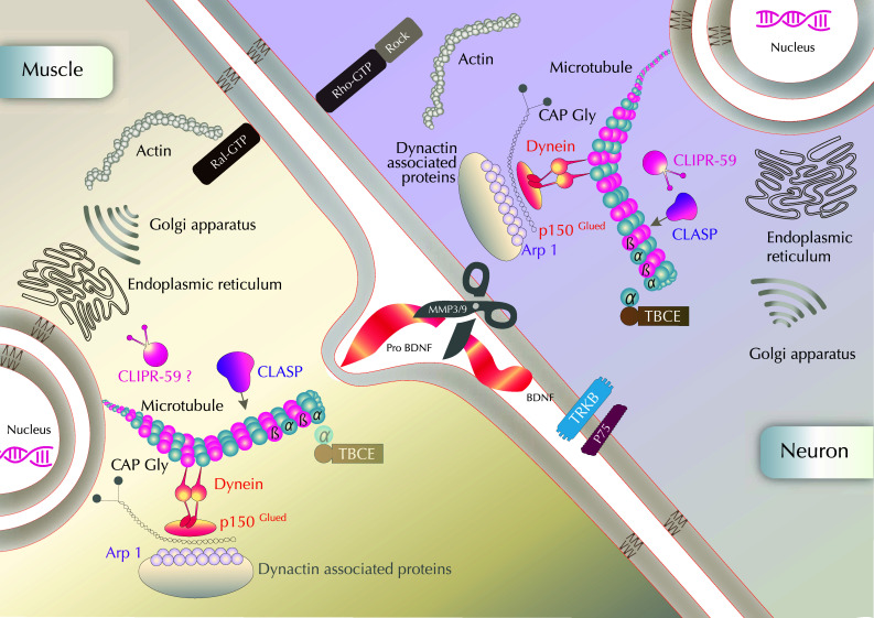
The neuromuscular junction (NMJ) is the synaptic connection between motor neurons and muscle fibers. It is involved in crucial processes such as body movements and breathing. Its proper development requires the guidance of motor axons toward their specific targets, the development of multi-innervated myofibers, and a selective synapse stabilization. It first consists of the removal of excessive motor axons on myofibers, going from multi-innervation to a single innervation of each myofiber. Whereas guidance cues of motor axons toward their specific muscular targets are well characterized, only few molecular and cellular cues have been reported as clues for selecting and stabilizing specific neuromuscular junctions. We will first provide a brief summary on NMJ development. We will then review molecular cues that are involved in NMJ stabilization, in both pre- and post-synaptic compartments, considering motor neurons and Schwann cells on the one hand, and muscle on the other hand. We will provide links with pathologies and highlight advances that can be brought both by basic research on NMJ development and clinical data resulting from the analyses of neurodegeneration of synaptic connections to obtain a better understanding of this process. The goal of this review is to highlight the findings toward understanding the roles of poly- or single-innervations and the underlying mechanisms of NMJ stabilization.


Conflict of interest statement

The authors declare no competing financial interests.

Figures

Fig. 1
Fig. 1
Critical periods of NMJ development with a focus on the elimination of multi-innervation. Motoneurons emit axons toward myotubes from E10 to E15 in mice. In parallel, Schwann cells migrate to reach the NMJ from E16 and cap the terminal. The differentiation of myoblasts into myotubes and muscle fibers is also schematized altogether with the time course. Dispersed acetylcholine receptors (AChRs) are expressed at moderate levels throughout myotubes surface prior to synapse formation, from E15 at the period of polyneuronal innervation, i.e. when each muscle fiber is still innervated by one or several axons of motor neurons (MNs). Postnatally, a critical step with the redrawal of about half multi-innervation occurs between P0 to P7, and the process of synapse elimination is achieved 2 weeks postnatally. AChR clustering occurs in the post-synaptic membrane, altogether with a progressive transition from multiple to single innervation of the NMJ
Fig. 2
Fig. 2
Activity dependent NMJ maintenance/stabilization. a Two motoneurons with synchronous activity are schematized. A similar activity will allow the synthesis of an equivalent amount of punishment (synaptotoxic factor) and reward (synaptotrophic factor) by both MNs, and their survival. b Inactive MNs will not be induced to produce either punishment or reward signal; the absence of competition for survival factor will allow the maintenance of both MNs. c The stabilization of one out of two MNs axons innervating a similar myofiber will be regulated by the activity: the active MN will synthesis both protective (reward) and punishment signals, that will allow its survival, whereas the inactive MN will receive punishment signals from the neighboring active MN only, that will lead to its elimination. Asynchronous AChR activation allows muscle to selectively destabilize synaptic sites to be eliminated
Fig. 3
Fig. 3
Molecular cues in NMJ formation/stabilization: a cross-talk between pre- and post-synaptic components. The agrin–muscle-specific kinase (MuSK)-rapsyn-AchR pathway is schematized. LRP4 acts as a co-factor for MuSK in agrin signaling. Agrin activates MuSK to cluster AChRs through the cytoplasmic linker protein rapsyn. Neuregulin that binds to ErbB receptors may also induce AChR transcription and agrin would direct AChR clustering. Neuregulin signaling also occurs from the axon to control Schwann cell survival. Schwann cells also belong to the tripartite NMJ with MN and muscle, and are essential for axon maintenance. Homophilic adhesion molecules such as NCAM are expressed on the surface of the three cell types composing the NMJ. Receptors to neurotrophic factors such as TrkB, p75 and GDNF receptors, are expressed at the MN surface. Actin regulators are present in the post-synaptic compartment, in particular Nogo-A, dystrophin and β-catenin. β-Catenin interacts with rapsyn and α-catenin to favor AChR clustering. β-Catenin-dependent transcription is also necessary for NMJ maintenance. Myogenin is involved in AchR expression, stabilization and clustering. Synaptic muscle fiber basal lamina is rich in laminin β2. It binds to and clusters the P/Q-type calcium channels that flank active zones and recruit other pre-synaptic components
Fig. 4
Fig. 4
Possible intracellular mechanisms of NMJ stabilization through a focus on actors interacting with cytoskeleton and organelles The dynamics and stability of both actin and microtubules regulate NMJ maintenance. Dynactin complex includes among others Arp1, p150glued and dyneins. Although present in both pre- and post-synaptic compartments, the TBCE protein accumulates at the Golgi apparatus and has been shown to be mainly required for maintenance of microtubules in distal axons so far. CLIPR-59 is located at the trans-Golgi network (TGN) and is proposed to affect protein/membrane trafficking or cytoskeleton remodeling at the NMJ, as well as in the pre-synaptic compartment, although its possible post-synaptic localization and role remain to be further analyzed. Molecular candidates have been proposed to act as synaptotoxic and synaptotrophic cues in NMJ stability: MMP3 and MMP9 located at the active terminals could cleave proBDNF at the active terminal during synaptic competition. The conversion of pro-brain-derived neurotrophic factor (proBDNF) to mature (m)BDNF would be activity-dependent and mediate synaptic competition and cell survival after endocytosis

References

    1. Gould TW, Burek MJ, Sosnowski JM, Prevette D, Oppenheim RW. The spatial-temporal gradient of naturally occurring motoneuron death reflects the time of prior exit from the cell cycle and position within the lateral motor column. Dev Biol. 1999;216:611–621. doi: 10.1006/dbio.1999.9490. - DOI - PubMed
    1. Tsuchida T, Ensini M, Morton SB, Baldassare M, Edlund T, Jessell TM, Pfaff SL. Topographic organization of embryonic motor neurons defined by expression of LIM homeobox genes. Cell. 1994;79:957–970. doi: 10.1016/0092-8674(94)90027-2. - DOI - PubMed
    1. Sharma K, Leonard AE, Lettieri K, Pfaff SL. Genetic and epigenetic mechanisms contribute to motor neuron pathfinding. Nature. 2000;406:515–519. doi: 10.1038/35020078. - DOI - PubMed
    1. Landmesser LT. The acquisition of motoneuron subtype identity and motor circuit formation. Int J Dev Neurosci. 2001;19:175–182. doi: 10.1016/S0736-5748(00)00090-3. - DOI - PubMed
    1. Kanning KC, Kaplan A, Henderson CE. Motor neuron diversity in development and disease. Annu Rev Neurosci. 2010;33:409–440. doi: 10.1146/annurev.neuro.051508.135722. - DOI - PubMed

Publication types