. 2015 Jan;45(1):87-97.
doi: 10.1183/09031936.00042414. Epub 2014 Oct 30.

Phenotype of asthmatics with increased airway S-nitrosoglutathione reductase activity

Affiliations

Phenotype of asthmatics with increased airway S-nitrosoglutathione reductase activity

Nadzeya V Marozkina et al. Eur Respir J. 2015 Jan.

Erratum in

  • Erratum. Phenotype of asthmatics with increased airway S-nitrosoglutathione reductase activity.
    Marozkina NV, Wang XQ, Stsiapura V, Fitzpatrick A, Carraro S, Hawkins GA, Bleecker E, Meyers D, Jarjour N, Fain SB, Wenzel S, Busse W, Castro M, Panettieri RA Jr, Moore W, Lewis SJ, Palmer LA, Altes T, de Lange EE, Erzurum S, Teague WG, Gaston B. Marozkina NV, et al. Eur Respir J. 2015 Jun;45(6):1763. doi: 10.1183/09031936.50042414. Eur Respir J. 2015. PMID: 26028625 No abstract available.

Abstract

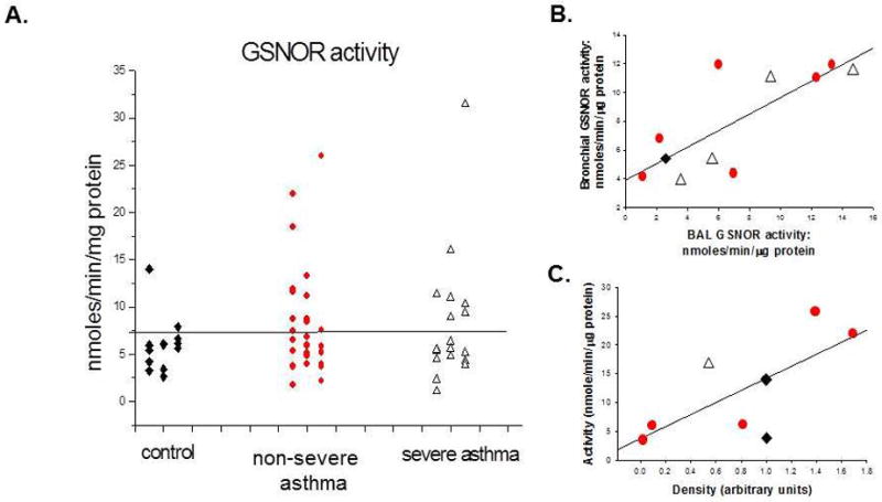
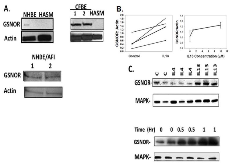
S-Nitrosoglutathione is an endogenous airway smooth muscle relaxant. Increased airway S-nitrosoglutathione breakdown occurs in some asthma patients. We asked whether patients with increased airway catabolism of this molecule had clinical features that distinguished them from other asthma patients. We measured S-nitrosoglutathione reductase expression and activity in bronchoscopy samples taken from 66 subjects in the Severe Asthma Research Program. We also analysed phenotype and genotype data taken from the program as a whole. Airway S-nitrosoglutathione reductase activity was increased in asthma patients (p=0.032). However, only a subpopulation was affected and this subpopulation was not defined by a "severe asthma" diagnosis. Subjects with increased activity were younger, had higher IgE and an earlier onset of symptoms. Consistent with a link between S-nitrosoglutathione biochemistry and atopy: 1) interleukin 13 increased S-nitrosoglutathione reductase expression and 2) subjects with an S-nitrosoglutathione reductase single nucleotide polymorphism previously associated with asthma had higher IgE than those without this single nucleotide polymorphism. Expression was higher in airway epithelium than in smooth muscle and was increased in regions of the asthmatic lung with decreased airflow. An early-onset, allergic phenotype characterises the asthma population with increased S-nitrosoglutathione reductase activity.


Figures

Figure 1
Figure 1. S-Nitrosoglutathione (GSNO) reductase activity in asthma
A. GSNO reductase activity was measured in the bronchoalveolar lavage (BAL) cell lysate of 54 subjects, including 13 control subjects, and 25 subjects with non-severe asthma and 16 subjects with severe asthma. Severe and non-severe asthma did not differ (p = ns). A greater proportion of subjects with asthma had high activity (>7.5 nmole/min/μg protein) than did controls (p = 0.032). B. A subset of subjects underwent analysis of an early (proximal) and late (distal) lavage fraction. Activities were weakly correlated between these two fractions (r2 = 0.61). C. Consistent with the previous report, activity was roughly correlated with protein expression (measured by immunoblot, densitometry) (r2 = 0.54). Activity was not correlated with BAL cell type (p = NS). Diamonds: controls; Circles: non-severe asthma; Triangles: severe asthma.
Figure 2
Figure 2. GSNO reductase expression is primarily epithelial cells
Lower lobe biopsy sections were embedded in paraffin and immunostained for S-Nitrosoglutathione reductase as described in the Methods. A. Control lung showing GSNO reductase immunoreactivity in the pseudostratified airway epithelium (100X). B. C. Non-severe asthma showing a similar distribution of GSNO reductase activity, particularly in the apex of the pseudostratified epithelium, less in basal membrane and smooth muscles. D.E. Severe asthma showing loss of epithelium and thickening of the basal membrane. Severe asthma (100X); note more prominent staining of the epithelium than of the smooth muscle. F. Control immunostaining of non-severe asthma showing absence of immunoreactivity in the absence of primary antibody.
Figure 3
Figure 3. GSNO reductase expression is greater in human airway epithelial cells than smooth muscle cells and is increased by interleukin (IL) 13
A. Immunoblots were performed on human normal bronchial epithelial cells (NHBE), both in monolayer culture and cultured at air-fluid interface (AFI). Studies were also performed on established cultures of human cystic fibrosis bronchial epithelial cells (CFBE) and on primary human airway smooth muscle (HASM) cells from six patients with asthma. Expression was greater in epithelial cells than in HASM cells, though there was variability in HASM expression. B. Left: freshly spun BAL cell pellets from severe asthma patients was exposed to interleukin IL 13 (10 ng/mL; 4 hr.) ex vivo. GSNO reductase expression was increased by interleukin-13 (n = 4; p < 0.05). Right: IL 13 incubation (2 hr.) also increased GSNO reductase expression in A549 cells in a dose-dependent manner. C. IL13, but not IL4, increased GSNO reductase expression (10 ng/mL each; 4 hr. p = 0.05 by ANOVA) relative to load control, and IL13 (10 ng/mL) also caused a time-dependent, 2-fold increase in GSNO reductase expression after 2 hours.
Figure 4
Figure 4. Regional inhomogeneity of inflammation and GSNO reductase expression in a severe asthma patient
On the day of hyperpolarized helium MRI scanning (21,27), the radiologist reported to the bronchoscopist the regions of good ventilation and poor ventilation to guide the biopsies. Green represents areas of good ventilation, and red represents poor ventilation. In A (coronal image) and B (transverse image) ventilation defects are shown at baseline in this stable, severe asthma patient. In C, a second image acquired after albuterol administration shows only moderate improvement in the left upper lobe ventilation defect. D and E. Inflammation and epithelial injury with increased GSNO reductase expression in the poorly ventilated lung unit (D, 40X; E, 100X). F. H&E staining in a sample from the poorly ventilated lung unit shows inflammation. G and H. GSNO reductase immunoreactivity in a sample from the well ventilated lung unit (G, 40X; H, 100X). I shows preserved airway epithelium, though thickened basement membrane, in a sample from the well ventilated lung unit (40X).
Figure 5
Figure 5. Proposed scheme showing the effect of increased GSNO reductase on asthma
A subset of patients, as previously reported (, –14) has a diathesis to have increased GSNO reductase expression. This is driven by allergic airway inflammation. In turn, this causes a decrease in airway GSNO levels that leads to smooth muscle constriction (–, –31) and further increase in Th2 cytokine expression (5).

References

    1. Gaston B, Reilly J, Drazen JM, Fackler J, Ramdev P, Arnelle D, Mullins ME, Sugarbaker DJ, Chee C, Singel DJ, Loscalzo J, Stamler JS. Endogenous nitrogen oxides and bronchodilator S-nitrosothiols in human airways. Proc Natl Acad Sci USA. 1993;90:10957–10961. - PMC - PubMed
    1. Gaston B, Drazen JM, Jansen A, Sugarbaker DA, Loscalzo J, Stamler JS. Relaxation of human bronchial smooth muscle by S-nitrosothiols in vitro. J Pharmacol Exp Ther. 1994;268:978–984. - PubMed
    1. Whalen EJ, Foster MW, Matsumoto A, Ozawa K, Violin JD, Que LG, Nelson CD, Benhar M, Keys JR, Rockman HA, Koch WJ, Daaka Y, Lefkowitz RJ, Stamler JS. Regulation of beta-adrenergic receptor signalling by S-nitrosylation of G-protein-coupled receptor kinase 2. Cell. 2007;129(3):511–522. - PubMed
    1. Gaston B, Doctor A, Singel D, Stamler JS. S-Nitrosothiol signalling in respiratory biology. Am J Respir Crit Care Med. 2006;173:1186–1193. - PMC - PubMed
    1. Foster M, Yang Z, Potts E, Foster W, Que L. S-nitrosoglutathione supplementation to ovalbumin-sensitized and -challenged mice ameliorates methacholine-induced bronchoconstriction. Am J Phyisol Lung Cell Mol Physiol. 2011;301:739–744. - PMC - PubMed

Publication types

MeSH terms

Substances