. 2014 Dec;5(6):657-66.
doi: 10.1007/s13244-014-0361-1. Epub 2014 Oct 31.

Multidetector computed tomography of mesenteric ischaemia

Affiliations

Multidetector computed tomography of mesenteric ischaemia

Andreu F Costa et al. Insights Imaging. 2014 Dec.

Abstract

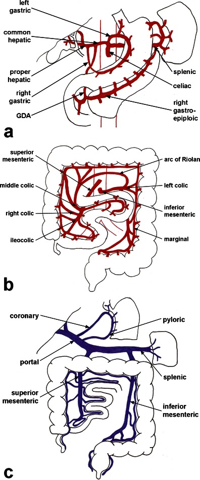
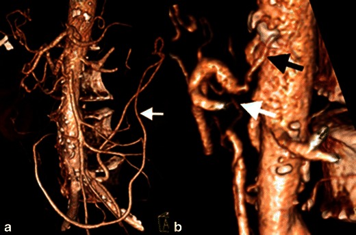
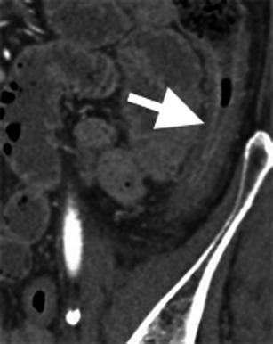
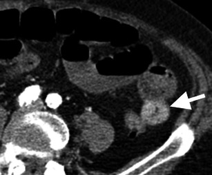
Mesenteric ischaemia comprises a broad, heterogeneous group of diseases characterised by inadequate blood supply to the small or large bowel. Acute mesenteric ischaemia is a surgical emergency, with significant associated morbidity and mortality. Because the clinical presentation of mesenteric ischaemia is variable and often nonspecific, a high index of clinical and radiologic suspicion is required for early diagnosis. The severity of mesenteric ischaemia ranges from transient, localised ischaemia to frank necrosis of the bowel. The most common causes of acute mesenteric ischaemia are embolic and thrombotic occlusion of the superior mesenteric artery, whereas chronic mesenteric ischaemia is almost always associated with generalised atherosclerotic disease. Multidetector computed tomography (MDCT) angiography is the preferred imaging test for acute and chronic mesenteric ischaemia. MDCT is useful in making a prompt, more precise diagnosis of mesenteric ischaemia, as well as identifying the cause and potential complications, which are key to reducing patient morbidity and mortality. In this article, we review the clinical features and aetiologies of mesenteric ischaemia and illustrate the imaging manifestations on MDCT.

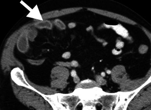
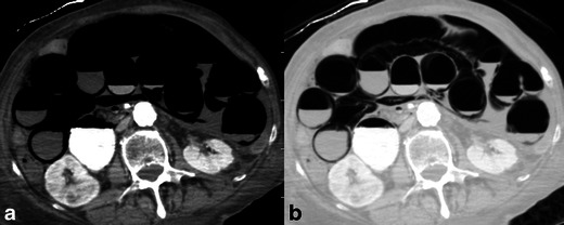
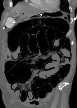
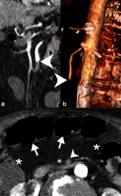
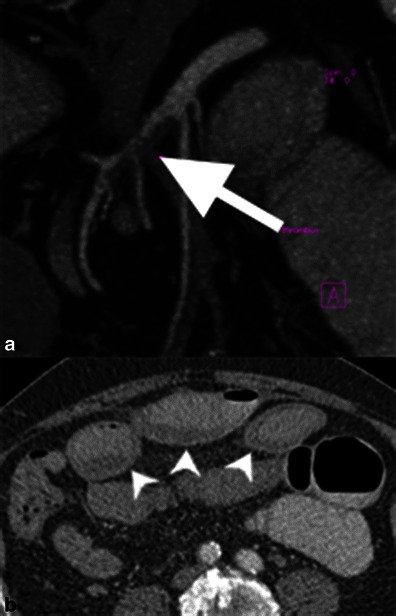
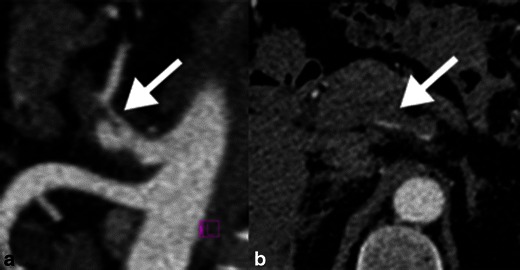
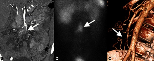
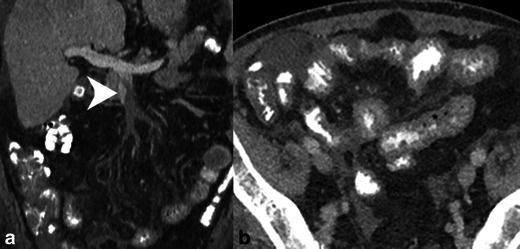
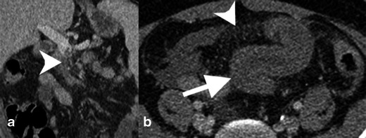
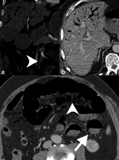
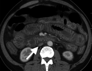
Main messages: • Acute and chronic mesenteric ischaemia are morbid conditions challenging to diagnose. • MDCT is the first-line imaging test for evaluating patients with suspected mesenteric ischaemia. • Bowel findings include wall thickening, abnormal enhancement, pneumatosis and luminal dilation. • Vascular occlusion, portomesenteric venous gas, mesenteric congestion and free air can be seen.


Figures

Fig. 1
Fig. 1
Mesenteric vasculature. Mesenteric arterial supply from the a coeliac and b superior and inferior mesenteric arteries. c Mesenteric venous drainage. GDA, gastroduodenal artery
Fig. 2
Fig. 2
Severe chronic mesenteric ischaemia. Patient with severe, three-vessel mesenteric atherosclerosis. The coeliac artery and superior mesenteric artery are occluded. a The inferior mesenteric artery is severely stenosed (>75 %) and filling via a prominent marginal artery of Drummond (white arrow). b Magnified view showing severe stenosis at the origin of the coeliac (black arrow) and superior mesenteric (white arrow) arteries
Fig. 3
Fig. 3
Bowel wall thickening in ischaemic colitis. Contrast-enhanced coronal reformatted image of a patient with severe chronic mesenteric ischaemia and diarrhoea. There is circumferential, oedematous wall thickening of the descending colon, pathologically proven to represent ischaemic colitis
Fig. 4
Fig. 4
Bowel wall thickening and haemorrhage. Axial CT image in a patient with an occluded superior mesenteric artery. A short segment of jejunum shows circumferential thickening and hyperattenuation, favoured to represent bowel wall heamatoma
Fig. 5
Fig. 5
Hyper-enhancement of the bowel mucosa. Axial contrast-enhanced CT image demonstrates hyper-enhancement of the right lower quadrant ileal mucosa (arrow) secondary to superior mesenteric artery occlusion (not shown). The degree of mucosal enhancement can be compared to left-sided bowel loops as an internal control
Fig. 6
Fig. 6
Pneumatosis intestinalis. Axial contrast-enhanced CT images in a soft tissue and b lung windows demonstrate extensive pneumatosis intestinalis secondary to superior mesenteric arterial occlusion. Note the increased conspicuity of pneumatosis with the use of lung windows
Fig. 7
Fig. 7
Dilated bowel in acute ischaemia. Coronal reformatted CT image demonstrates diffuse small bowel dilation with pneumatosis in a patient with extensive small bowel ischaemia. Portal venous gas in the liver is also evident
Fig. 8
Fig. 8
Embolic occlusion of the superior mesenteric artery. a Contrast-enhanced coronal reformatted CT image and b 3D volume-rendered reconstruction of demonstrate an abrupt cutoff of the proximal superior mesenteric artery (white arrowheads) secondary to an embolus. c Axial contrast-enhanced CT demonstrates relatively poor enhancement of the transverse colon (white arrows) relative to the hepatic and splenic flexures (*). Note also the occluded SMA (white arrowhead)
Fig. 9
Fig. 9
SMA thrombosis. a Contrast-enhanced, coronal reformatted CT images demonstrate an eccentric filling defect in the proximal SMA, corresponding to thrombus formation. b Axial contrast-enhanced CT image shows oedematous bowel wall thickening of small bowel loops (white arrowheads) and mesenteric congestion. This patient subsequently improved with conservative anticoagulation therapy
Fig. 10
Fig. 10
Spontaneous coeliac artery dissection. a Contrast-enhanced sagittal reformatted image demonstrates a dissection flap of the coeliac artery (white arrow). b Axial CT image shows the dissection flap continues into the common hepatic artery with thrombosis of the false lumen. There was no evidence of bowel ischaemia in this patient and the appearance remained stable on follow-up imaging
Fig. 11
Fig. 11
Mesenteric ischaemia secondary to vascular compression by a small bowel carcinoid. a Coronal contrast-enhanced reformatted image shows mesenteric metastatic disease from the small bowel carcinoid encasing distal branches of the SMA (white arrow). b Coronal image from an octreotide scan shows uptake by the metastatic disease (white arrow), indicating tumoral somatostatin receptors. c Three-dimensional volume-rendered reconstruction shows attenuated calibre of the ileocolic artery (white arrow)
Fig. 12
Fig. 12
SMV thrombosis. a Contrast-enhanced coronal reformatted CT demonstrates occlusive thrombus in the SMV near the portal confluence (white arrowhead). b Axial CT image demonstrates diffuse small bowel wall thickening secondary to venous congestion and ischaemia
Fig. 13
Fig. 13
SMV thrombosis with mesenteric congestion. a Contrast-enhanced coronal reformatted CT of an occlusive thrombus in the SMV (white arrowhead) extending to the portal confluence. b Axial contrast-enhanced CT image demonstrates mesenteric congestion with engorged mesenteric veins and trace fluid (white arrowhead). The small bowel is thickened because of the venous stasis and probable ischaemia
Fig. 14
Fig. 14
Portomesenteric venous gas. a Contrast-enhanced coronal reformatted CT demonstrates occlusion of the SMA (arrowhead). Portal venous gas is seen in the liver. b Axial CT shows portal venous gas in the anti-dependent liver. c The small bowel demonstrates luminal dilation, a paper-thin wall and poorly enhancing mucosa. There is mesenteric venous gas (arrowhead) as well as pneumatosis in the dependent bowel wall. The patient had established bowel necrosis and died shortly after imaging
Fig. 15
Fig. 15
Shock bowel. Contrast-enhanced axial CT image demonstrates diffuse bowel wall thickening with mural hyper-enhancement. Note the secondary sign of a flattened inferior vena cava, indicating hypovolaemia

References

    1. Herbert GS, Steele SR. Acute and chronic mesenteric ischemia. Surg Clin N Am. 2007;87(5):1115–1134. doi: 10.1016/j.suc.2007.07.016. - DOI - PubMed
    1. Gore RM, Yaghmai V, Thakrar KH, Berlin JW, Mehta UK, Newmark GM, et al. Imaging in intestinal ischemic disorders. Radiol Clin N Am. 2008;46(5):845–875. doi: 10.1016/j.rcl.2008.05.004. - DOI - PubMed
    1. Levy AD. Mesenteric ischemia. Radiol Clin N Am. 2007;45(3):593–599. doi: 10.1016/j.rcl.2007.04.012. - DOI - PubMed
    1. Bobadilla JL. Mesenteric ischemia. Surg Clin N Am. 2013;93(4):925–940. doi: 10.1016/j.suc.2013.04.002. - DOI - PubMed
    1. Sreenarasimhaiah J. Chronic mesenteric ischemia. Best Pract Res Clin Gastroenterol. 2005;19(2):283–295. doi: 10.1016/j.bpg.2004.11.002. - DOI - PubMed

LinkOut - more resources