. 2014 Dec;141(23):4479-88.
doi: 10.1242/dev.116467. Epub 2014 Oct 30.

Adipocyte amino acid sensing controls adult germline stem cell number via the amino acid response pathway and independently of Target of Rapamycin signaling in Drosophila

Affiliations

Adipocyte amino acid sensing controls adult germline stem cell number via the amino acid response pathway and independently of Target of Rapamycin signaling in Drosophila

Alissa R Armstrong et al. Development. 2014 Dec.

Abstract

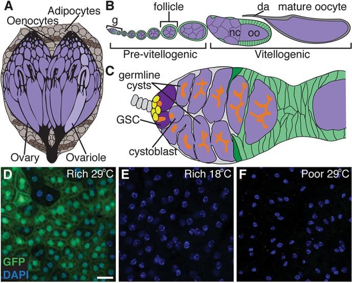
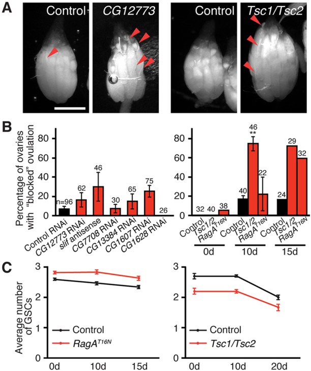
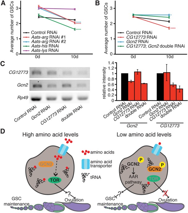
How adipocytes contribute to the physiological control of stem cells is a critical question towards understanding the link between obesity and multiple diseases, including cancers. Previous studies have revealed that adult stem cells are influenced by whole-body physiology through multiple diet-dependent factors. For example, nutrient-dependent pathways acting within the Drosophila ovary control the number and proliferation of germline stem cells (GSCs). The potential role of nutrient sensing by adipocytes in modulating stem cells in other organs, however, remains largely unexplored. Here, we report that amino acid sensing by adult adipocytes specifically modulates the maintenance of GSCs through a Target of Rapamycin-independent mechanism. Instead, reduced amino acid levels and the consequent increase in uncoupled tRNAs trigger activation of the GCN2-dependent amino acid response pathway within adipocytes, causing increased rates of GSC loss. These studies reveal a new step in adipocyte-stem cell crosstalk.

Keywords: Adipocytes; Amino acid transporters; Diet; Drosophila; Germline stem cells; Oogenesis.


Figures

Fig. 1.
Fig. 1.
A tool to determine how genetic manipulation of nutrient-dependent pathways in adult adipocytes impacts the GSC lineage in the Drosophila ovary. (A) The Drosophila fat body is an endocrine organ awash in hemolymph and composed of sheets of adipocytes intercalated with hepatocyte-like oenocytes. The fat body underlies the cuticle and surrounds the brain, gut and ovaries in females. (B) Developing follicles arranged in chronological order make up an ovariole. Follicles, formed in an anterior germarium (g), are germline cysts (one oocyte, oo, plus 15 nurse cells, nc; purple) surrounded by follicle cells (green), and develop to form a mature oocyte containing a dorsal appendage (da). (C) Each germarium contains two or three GSCs in a well-defined niche composed primarily of cap cells (yellow), and each GSC division yields a GSC and a cystoblast that forms a 16-cell cyst. GSCs and other early germline stages are identifiable based on the position and morphology of a germline-specific organelle, the fusome (orange). Follicle cells derived from follicle stem cells (dark green) envelop the cyst, making a follicle. (D-F) In females raised at 18°C and subsequently switched to 29°C, Gal80ts; Lsp2 drives UAS transgene expression specifically in adult adipocytes (see supplementary material Figs S1 and S2). A UAS-GFP reporter (green) driven by Gal80ts; Lsp2 shows robust expression in adipocytes on a rich diet at 29°C (D), but is not expressed either at 18°C (E) or on a poor diet (F). DAPI (blue) labels nuclei. Scale bar: 50 μm.
Fig. 2.
Fig. 2.
Adult adipocyte-specific knockdown of amino acid transporters does not cause obvious changes in ovarian or adipocyte morphology. (A,B) RT-PCR analysis of hand-dissected fat bodies showing knockdown of amino acid transporters (A), and normal ovariole and adipocyte morphology (B) after 10 days of Gal80ts; Lsp2-mediated induction of RNAi or antisense transgenes against amino acid transporters or white control. Rp49 is a control. DAPI (white or blue) labels nuclei. In adipocytes, Nile Red, which fluoresces in both red and green (Greenspan et al., 1985), is shown in green. Scale bars: 100 μm (ovarioles); 10 μm (adipocytes).
Fig. 3.
Fig. 3.
Adult adipocyte-specific knockdown of a subset of amino acid transporters encoded by slif, CG7708, CG1607, CG13384, CG1628 and CG12773 results in reduced egg production. (A) Females carrying Gal80ts; Lsp2 and UAS-RNAi transgenes against amino acid transporters [or a UAS-antisense transgene (Colombani et al., 2003) in the case of slif] or a control UAS-RNAi (against white, an eye color gene) raised at 18°C and switched to 29°C for adult adipocyte-specific knockdown for the indicated number of days. Knockdown of a subset of transporters causes a significant decrease in the average number of eggs laid per female per day. Transporters in red font were followed up on in this study. (B) Control females carrying UAS-RNAi or -antisense transgenes against amino acid transporters in the absence of Gal80ts; Lsp2 and subjected to the same temperature regime as in A show egg-laying rates that are statistically indistinguishable from those of a UAS-GFPRNAi control, except for UAS-CG13384RNAi. *P<0.05; **P<0.01; ***P<0.001, Student's t-test. Data shown as mean±s.e.m.
Fig. 4.
Fig. 4.
Adult adipocyte-specific knockdown of amino acid transporters leads to increased rates of GSC loss in the ovary. (A) Average number of GSCs per germarium at 0, 5 or 10 days of Gal80ts; Lsp2-mediated induction of RNAi or antisense transgenes against amino acid transporters or white control. See supplementary material Fig. S3 for sample sizes and distribution. **P<0.01; ****P<0.0001, two-way ANOVA with interaction. Data shown as mean±s.e.m. (B) Germaria at 10 days of adipocyte-specific GFP control or amino acid transporter RNAi labeled for phosphorylated Mad (pMad; green), 1B1 (red, fusome) and Lamin C (red, cap cell nuclear envelope). GSC nuclei are outlined. Scale bar: 2.5 μm. (C) Box and whisker plot of mean nuclear pMad intensity for experiment in B. Sample sizes are included above. **P<0.01; ***P<0.001, Student's t-test.
Fig. 5.
Fig. 5.
Reduced amino acid transport in adipocytes does not affect cell death within the germarium, but causes a slight increase in GSC proliferation. (A) Germaria from females at 10 days of adult adipocyte-specific knockdown of CG1628 or white control showing some occurrence of cell death (ApopTag, green) in both cases. DAPI (blue) labels nuclei; 1B1 (red) labels fusomes; LamC (red) labels cap cell nuclear envelopes. Scale bar: 10 μm. In the graph on right, bars represent the percentage of germaria containing ApopTag-positive cells, with the hatched region indicating the fraction of those displaying ApopTag adjacent to GSC niche. The number of germaria analyzed is shown above each bar. (B,C) Frequencies of GSCs (B) or follicle cells (C) in S phase, based on EdU incorporation, at 10 days of adipocyte knockdown of amino acid transporters or GFP control. Number of GSCs (B) or follicle cell fields (C) analyzed is shown above each bar. *P<0.05; **P<0.01; ***P<0.001, Student's t-test. Data shown as mean±s.e.m.
Fig. 6.
Fig. 6.
GSC loss induced by adult adipocyte-specific knockdown of amino acid transporters is independent of TOR signaling. (A) Ovaries at 10 days of adipocyte-specific CG12773 knockdown or Tsc1/Tsc2 induction showing retention of mature oocytes in subsets of ovarioles (‘blocked’ ovulation). Mature oocytes are recognizable by the presence of dorsal appendages (arrowheads). Scale bar: 500 μm. (B) Percentage of ovaries containing at least one ovariole that retains more than one mature oocyte at 10 days of adipocyte-specific amino acid transporter knockdown (left) or at different days of inhibition of TOR signaling (right). **P<0.01. Data from 0 d and 15 d time points are from one experiment, whereas 10 d represents three experiments. Number of ovaries analyzed is shown above each bar. (C) Average number of GSCs at different days of Gal80ts; Lsp2-mediated induction of a dominant-negative RagA (RagAT16N) or of Tsc1/Tsc2 transgenes showing that inhibition of TOR signaling has no effect on GSC maintenance. See supplementary material Fig. S6 for sample sizes and distribution. Control is GFP RNAi for A and B (left), and Gal80ts; Lsp2 alone for B (right) and C.
Fig. 7.
Fig. 7.
The amino acid response pathway within adipocytes contributes to the control of GSC maintenance. (A,B) Average number of GSCs at 0 or 10 days of Gal80ts; Lsp2-mediated induction of GFP control, Arginyl-tRNA synthetase (Aats-arg), Histidyl-tRNA synthetase (Aats-his), Lysyl-tRNA synthetase (Aats-lys), CG12773, Gcn2 and double CG12773 Gcn2 RNAi transgenes. See supplementary material Fig. S7 for sample sizes and distribution, and for efficiency of knockdown for experiment in A. Numbers of germaria analyzed in B are: 131 for control 0 d; 71 for control 10 d; 121 for CG12773 0d; 89 for CG12773; 121 for Gcn2 0 d; 102 for Gcn2 10 d; 159 for CG12773 Gcn2 double 0 d; 90 for CG12773 Gcn2 double 10 d. *P<0.05; ****P≤0.0001, two-way ANOVA with interaction. Data are mean±s.e.m. (C) RT-PCR analysis of hand-dissected fat bodies showing specific knockdown of Gcn2 and/or CG12773 in genotypes shown in B. Data shown as mean±s.e.m. (D) Model for how amino acid sensing within adipocytes regulates the GSC lineage. Under high amino acid levels, the amino acid response (AAR) pathway is off and TOR is active, resulting in optimal GSC maintenance and ovulation rates. Under lower amino acid levels, the AAR pathway is triggered through an increase in unloaded tRNAs and activation of GCN2 kinase, leading to GSC loss. Reduced TOR activity causes a partial block in ovulation.

References

    1. Ables E. T. and Drummond-Barbosa D. (2010). The steroid hormone ecdysone functions with intrinsic chromatin remodeling factors to control female germline stem cells in Drosophila. Cell Stem Cell 7, 581-592 10.1016/j.stem.2010.10.001 - DOI - PMC - PubMed
    1. Ables E. T., Laws K. M. and Drummond-Barbosa D. (2012). Control of adult stem cells in vivo by a dynamic physiological environment: diet-dependent systemic factors in Drosophila and beyond. Wiley Interdiscip. Rev. Dev. Biol. 1, 657-674 10.1002/wdev.48 - DOI - PMC - PubMed
    1. Arquier N., Géminard C., Bourouis M., Jarretou G., Honegger B., Paix A. and Léopold P. (2008). Drosophila ALS regulates growth and metabolism through functional interaction with insulin-like peptides. Cell Metab. 7, 333-338 10.1016/j.cmet.2008.02.003 - DOI - PubMed
    1. Arrese E. L. and Soulages J. L. (2010). Insect fat body: energy, metabolism, and regulation. Annu. Rev. Entomol. 55, 207-225 10.1146/annurev-ento-112408-085356 - DOI - PMC - PubMed
    1. Bianchini F., Kaaks R. and Vainio H. (2002). Overweight, obesity, and cancer risk. Lancet Oncol. 3, 565-574 10.1016/S1470-2045(02)00849-5 - DOI - PubMed

Publication types

LinkOut - more resources