. 2014 Dec 19;289(51):35656-67.
doi: 10.1074/jbc.M114.595082. Epub 2014 Oct 30.

The role of the strictly conserved positively charged residue differs among the Gram-positive, Gram-negative, and chloroplast YidC homologs

Affiliations

The role of the strictly conserved positively charged residue differs among the Gram-positive, Gram-negative, and chloroplast YidC homologs

Yuanyuan Chen et al. J Biol Chem. .

Abstract

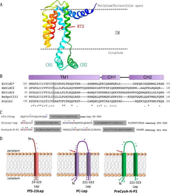
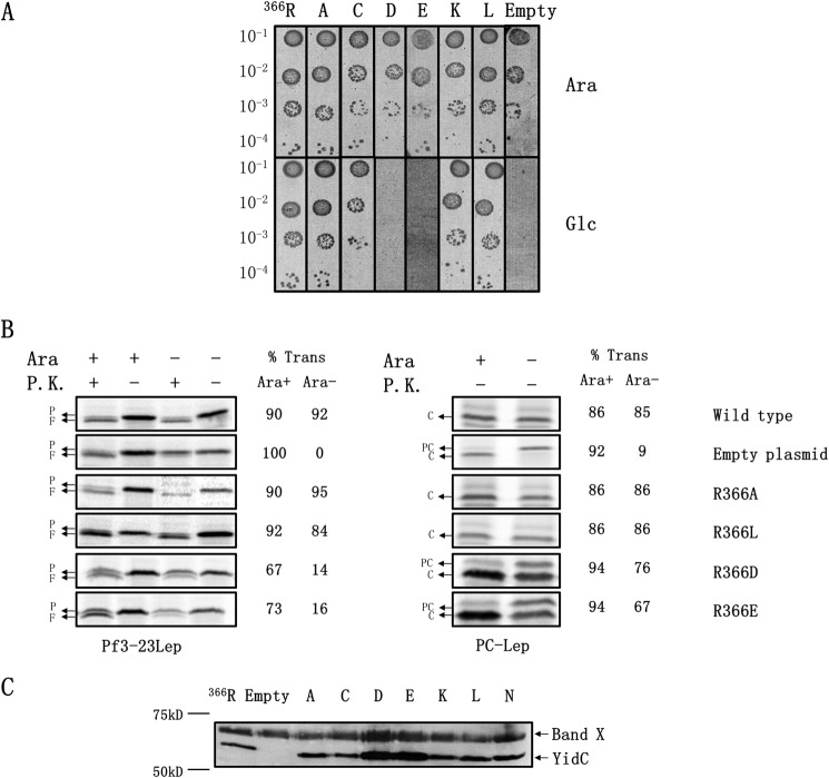
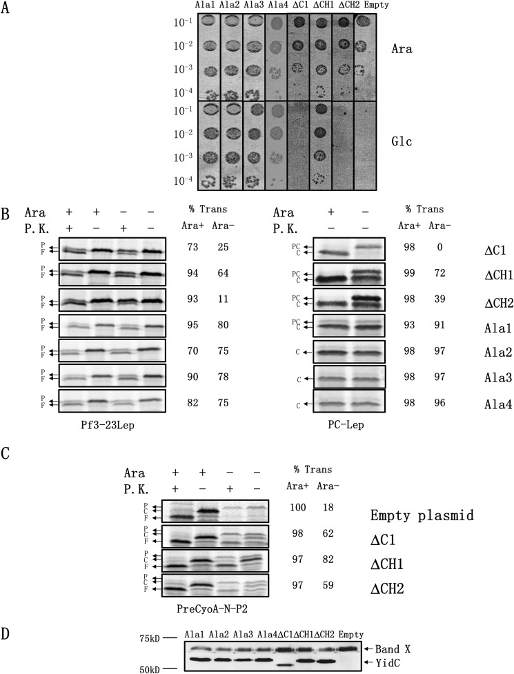
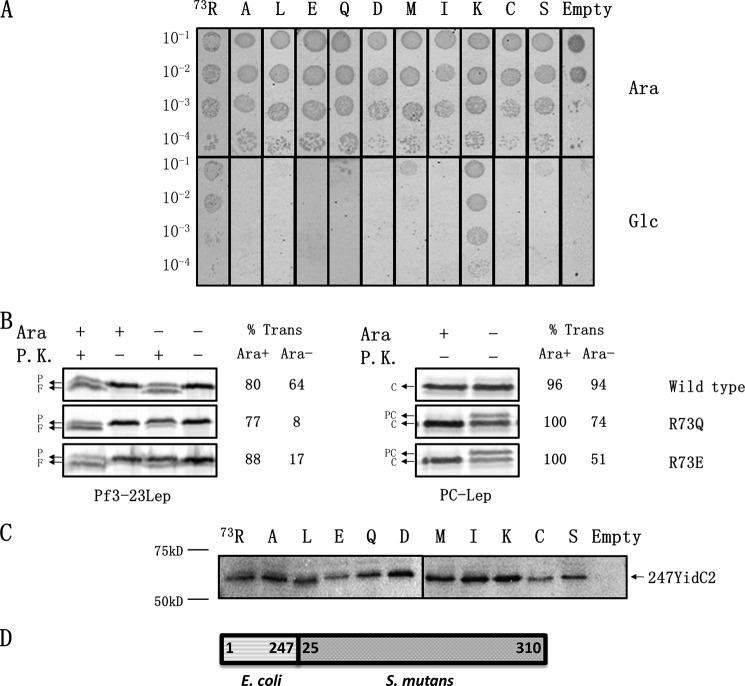
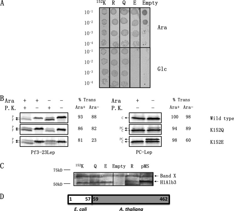
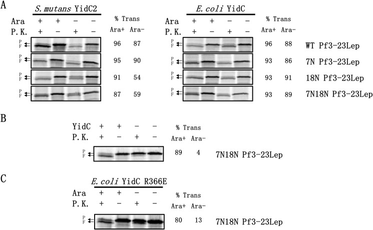
Recently, the structure of YidC2 from Bacillus halodurans revealed that the conserved positively charged residue within transmembrane segment one (at position 72) is located in a hydrophilic groove that is embedded in the inner leaflet of the lipid bilayer. The arginine residue was essential for the Bacillus subtilis SpoIIIJ (YidC1) to insert MifM and to complement a SpoIIIJ mutant strain. Here, we investigated the importance of the conserved positively charged residue for the function of the Escherichia coli YidC, Streptococcus mutans YidC2, and the chloroplast Arabidopsis thaliana Alb3. Like the Gram-positive B. subtilis SpoIIIJ, the conserved arginine was required for functioning of the Gram-positive S. mutans YidC2 and was necessary to complement the E. coli YidC depletion strain and to promote insertion of a YidC-dependent membrane protein synthesized with one but not two hydrophobic segments. In contrast, the conserved positively charged residue was not required for the E. coli YidC or the A. thaliana Alb3 to functionally complement the E. coli YidC depletion strain or to promote insertion of YidC-dependent membrane proteins. Our results also show that the C-terminal half of the helical hairpin structure in cytoplasmic loop C1 is important for the activity of YidC because various deletions in the region either eliminate or impair YidC function. The results here underscore the importance of the cytoplasmic hairpin region for YidC and show that the arginine is critical for the tested Gram-positive YidC homolog but is not essential for the tested Gram-negative and chloroplast YidC homologs.

Keywords: Alb3; Membrane Biogenesis; Membrane Enzyme; Membrane Function; Membrane Insertion; Membrane Protein; Membrane Structure; Structure-Function; YidC; YidC Mechanism.


Figures

FIGURE 1.
FIGURE 1.
Crystal structure of B. halodurans YidC2 highlighting the conserved positive charge in the hydrophilic groove. A, ribbon representation of the structure of the B. halodurans YidC2. The conserved positively charged Arg-72 (corresponding to Arg-366 in E. coli YidC) is highlighted in red and is located near the top of the hydrophilic groove. B, sequence alignment between YidC homologs in E. coli (Ec), S. mutans (Sm), B. halodurans (Bh), B. subtilis (Bs), and A. thaliana (At). The conserved positively charged residue in TM1 (corresponding to TM2 in the E. coli YidC) that is essential for function of the B. subtilis SpoIIIJ (YidC1) is highlighted in gray. CH1 and CH2 correspond to the N- and C-terminal amino acid sequences that make up the helical hairpin structure. C, YidC substrate constructs used in this study (for details see “Experimental Procedures”), and D, topology of the substrates Pf3-23Lep, PC-Lep, and PreCyoA-N-P2. The red arrows highlight the cleavage site for signal peptidases.
FIGURE 2.
FIGURE 2.
Arginine 366 in the E. coli YidC is dispensable for function. A, complementation assay to test the importance of Arg-366 for E. coli YidC function. Arg-366 within the E. coli YidC was mutated to Ala, Cys, Asp, Glu, Lys, and Leu in pACYC184. The pACYC184-encoded YidC mutants were transformed and expressed in E. coli JS7131 under YidC depletion conditions for 3 h. Expression of the plasmid-encoded YidC mutants was under the endogenous yidC promoter. Serial dilutions of the cultures were spotted and incubated under wild-type YidC expression conditions (top panels) and depletion conditions (lower panels), respectively. Plasmid-encoded wild-type YidC was also expressed, as a positive control, to show it was capable of supporting growth of the cell when chromosomal YidC was depleted. B, E. coli YidC neutral and negatively charged mutants at 366 were tested for their ability to promote the insertion of Pf3-23Lep and PC-Lep. JS7131 encoding the pACYC184-YidC wild-type, mutants, or the pACYC184 empty vector were grown for 3 h under YidC expression (0.2% arabinose) or YidC depletion conditions (0.2% glucose). The respective strains were co-transformed with pMS119 encoding Pf3-23Lep or PC-Lep. Expression of the YidC substrate was induced by the addition of 1 mm isopropyl β-d-1-thiogalactopyranoside, and the cells were labeled with [35S]methionine for 1 min. Protease accessibility assay (left panels) was used to monitor translocation of the N-tail of Pf3-23Lep. After [35S] labeling, the cells were converted to spheroplasts, and a portion was treated with proteinase K (P.K.), as described under “Experimental Procedures.” Signal peptide processing (right panels) was employed to measure membrane insertion of PC-Lep. After labeling with [35S]methionine for 1 min, the cells were precipitated with TCA and analyzed as described under “Experimental Procedures.” The percent translocation of Pf3-23Lep and PC-Lep was quantified as described under “Experimental Procedures.” P represents Pf3-23Lep protein; F denotes the proteinase K fragment; PC represents PC-Lep; C denotes C-Lep protein generated by SP1 cleavage. C, Western blot to examine expression and stability of the E. coli YidC mutants using antiserum against a C-terminal peptide of EcYidC. The top band is an unrelated 70-kDa protein recognized by our antiserum. P.K., proteinase K.
FIGURE 3.
FIGURE 3.
C-terminal region of the helix hairpin region is important for the function of the E. coli YidC, but its precise sequence is not. A, complementation assay to examine the role of the helical hairpin structure for YidC function. The YidC depletion strain JS7131 was transformed with pACYC184-encoding YidC Ala-1, Ala-2, Ala-3, Ala-4, Δ371–416 (ΔC1), Δ389–398 (ΔCH1), and Δ399–415 (ΔCH2) mutants, and a spot test was used to examine complementation, as described in Fig. 2A. B, YidC hairpin mutants were tested for their ability to insert Pf3-23Lep and PC-Lep. JS7131 bearing pACYC184 encoding YidC ΔC1, ΔCH1, ΔCH2, Ala-1, Ala-2, Ala-3, or Ala-4 was transformed with either pMS119 Pf3-23Lep or PC-Lep. Expression of the YidC substrate, labeling, and membrane insertion was performed as described in Fig. 2B. Left panel shows protease mapping of Pf3-23Lep. Right panel shows signal peptide processing of PC-Lep. P represents Pf3-23Lep protein; F denotes the proteinase K fragment; PC represents PC-Lep; C denotes C-Lep protein generated by SP1 cleavage. C, YidC mutants with deletions in the C1 loop were tested for their ability to insert preCyoA-N-P2, as described in 2B. P corresponds to PreCyoA-N-P2; C denotes the CyoA-N-P2 produced by SP1 cleavage; F corresponds to the proteinase K fragment of CyoA-N-P2. D, Western blotting to detect expression of the E. coli YidC mutants. The percent translocation of Pf3-23Lep, PC-Lep, and PreCyoA-N-P2 was quantified as described under “Experimental Procedures.” P.K., proteinase K.
FIGURE 4.
FIGURE 4.
Positively charged residue at position 73 in the S. mutans YidC2 is critical for function. A, complementation assay to elucidate the importance of the conserved arginine for S. mutans 247YidC2 function. JS7131 bearing pACYC184 possessing the S. mutans 247YidC2 WT or mutants was tested for complementation using the spot test, as described in Fig. 2A. 247YidC2 was constructed by fusing residues 1–247 of the E. coli YidC to residues 25–310 of S. mutans YidC. Mutants studied are WT (73R), R73A, R73L, R73E, R73Q, R73D, R73M, R73I, R73K, R73C, and R73S. B, wild-type and representative mutants of 247YidC2, R73Q, and R73E were tested for their ability to insert Pf3-23Lep and PC-Lep into the membrane. Expression of YidC substrates, [35S] labeling, protease accessibility of Pf3-23Lep (left panels), and signal peptide cleavage of PC-Lep (right panels) was performed, as described in Fig. 2B. The percent translocation of Pf3-23Lep and PC-Lep was quantified as described under “Experimental Procedures.” P represents Pf3-23Lep protein; F denotes the proteinase K fragment; PC represents PC-Lep; C denotes C-Lep protein generated by SP1 cleavage. C, Western blot to examine expression levels of wild-type and 247YidC2 mutants using antibodies prepared against a C-terminal peptide of the S. mutans YidC2. D, schematic of S. mutans 247YidC2. It contains residues 1–247 of the E. coli YidC2 fused to residues 25–310 of S. mutans YidC2. P.K., proteinase K.
FIGURE 5.
FIGURE 5.
Positively charged residue at 152 in the A. thaliana Alb3 is not critical for function. A, complementation assay to determine the importance of the strictly conserved positively charged residue. The H1Alb3 WT, K152R, K152Q, and K152E mutants were tested for their ability to complement the YidC depletion strain using the spot test, as described in Fig. 2A. H1Alb3 contains the first 57 amino acids of E. coli YidC fused to residues 59–462 of Alb3. B, WT H1Alb3, K152Q and K152E were tested for their ability to insert Pf3-23Lep and PC-Lep using protease mapping (left panel) and signal peptide cleavage (right panel), respectively. The percent translocation of Pf3-23Lep and PC-Lep was quantified as described under “Experimental Procedures.” P represents Pf3-23Lep protein; F denotes the proteinase K fragment; PC represents PC-Lep; C denotes C-Lep protein generated by SP1 cleavage. C, Western blotting to examine expression of the wild-type and H1Alb3 mutants using antibodies generated against a C-terminal peptide from P. sativum Alb3. The top band is an unrelated band seen in our Western blots. The pMS lane refers to H1Alb3 overexpressed in pMS119 to confirm that the band we are examining is indeed H1Alb3. D, schematic of H1Alb3. It contains the first 57 amino acids of E. coli YidC, a linker valine residue, and residues 59–462 of Alb3. P.K., proteinase K.
FIGURE 6.
FIGURE 6.
Test of the electrostatic attraction model for how YidC facilitates translocation of the N-tail of Pf3–23Lep across the membrane. The negatively charged residue in the N-tail is important for membrane insertion mediated by S. mutans 247YidC2 but not by the E. coli YidC. A, E. coli YidC (right panel) or S. mutans YidC2 (left panel) was tested for their ability to insert Pf3-23Lep WT, Pf3-23Lep7N, Pf3-23Lep18N, and Pf3-23Lep7N18N. JS7131 bearing pACYC184 encoding E. coli YidC or S. mutans 247YidC2 was transformed with pMS119 encoding Pf3-23Lep proteins. Expression of the Pf3-23Lep mutants, labeling, and membrane insertion was performed as described in Fig. 2B. B, YidC is required for membrane insertion of Pf3-23Lep7N18N. JS7131 expressing YidC or depleted for YidC (−) was analyzed for N-tail translocation. P represents Pf3-23Lep protein; F denotes the proteinase K fragment. C, R366E YidC is defective in translocating the N-tail of Pf3-23Lep lacking negatively charged residues. JS7131 bearing pACYC184 encoding E. coli YidC R366E was transformed with pMS119 encoding Pf3-23Lep7N18N and analyzed for N-tail translocation, as described in Fig. 2B. The percent translocation of Pf3-23Lep and PC-Lep was quantified as described under “Experimental Procedures.” P.K., proteinase K.

References

    1. Wang P., Dalbey R. E. (2011) Inserting membrane proteins: the YidC/Oxa1/Alb3 machinery in bacteria, mitochondria, and chloroplasts. Biochim. Biophys. Acta 1808, 866–875 - PubMed
    1. Kiefer D., Kuhn A. (2007) YidC as an essential and multifunctional component in membrane protein assembly. Int. Rev. Cytol. 259, 113–138 - PubMed
    1. Dalbey R. E., Kuhn A., Zhu L., Kiefer D. (2014) The membrane insertase YidC. Biochim. Biophys. Acta 1843, 1489–1496 - PubMed
    1. Scotti P. A., Urbanus M. L., Brunner J., de Gier J. W., von Heijne G., van der Does C., Driessen A. J., Oudega B., Luirink J. (2000) YidC, the Escherichia coli homologue of mitochondrial Oxa1p, is a component of the Sec translocase. EMBO J. 19, 542–549 - PMC - PubMed
    1. Serek J., Bauer-Manz G., Struhalla G., van den Berg L., Kiefer D., Dalbey R. E., Kuhn A. (2004) Escherichia coli YidC is a membrane insertase for Sec-independent proteins. EMBO J. 23, 294–301 - PMC - PubMed

Publication types

MeSH terms