. 2015 Jan 15;125(3):562-9.
doi: 10.1182/blood-2014-07-589507. Epub 2014 Oct 30.

Identification of a juxtamembrane mechanosensitive domain in the platelet mechanosensor glycoprotein Ib-IX complex

Affiliations

Identification of a juxtamembrane mechanosensitive domain in the platelet mechanosensor glycoprotein Ib-IX complex

Wei Zhang et al. Blood. .

Abstract

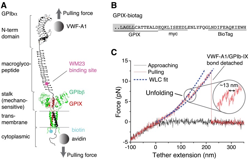
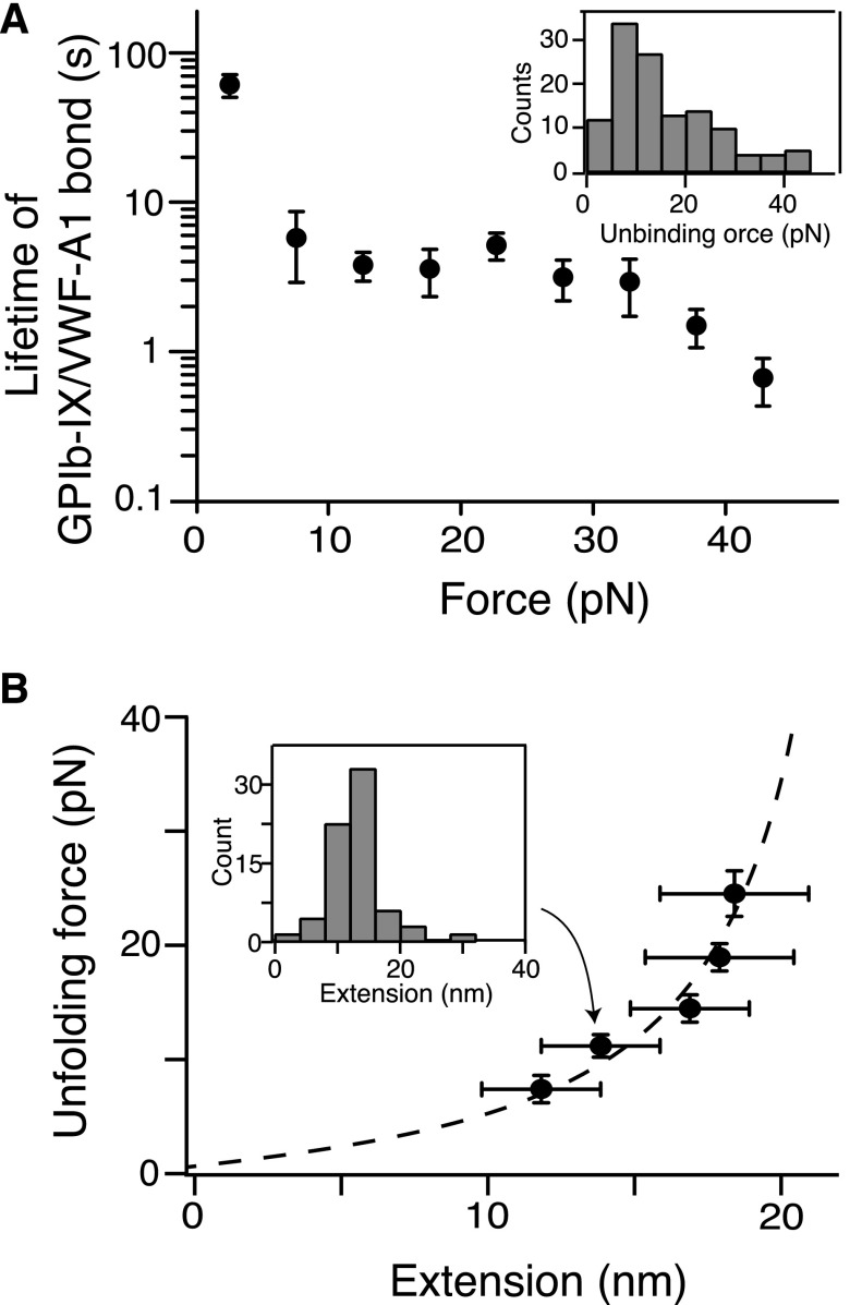
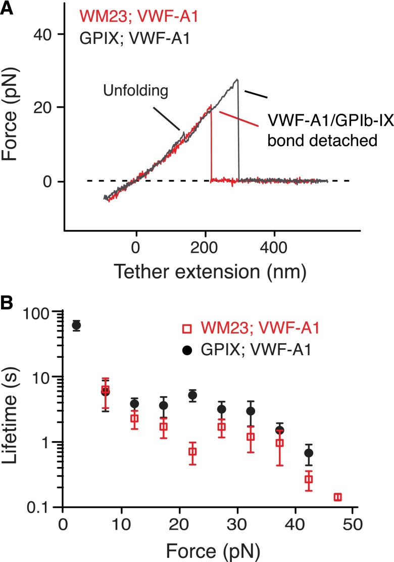
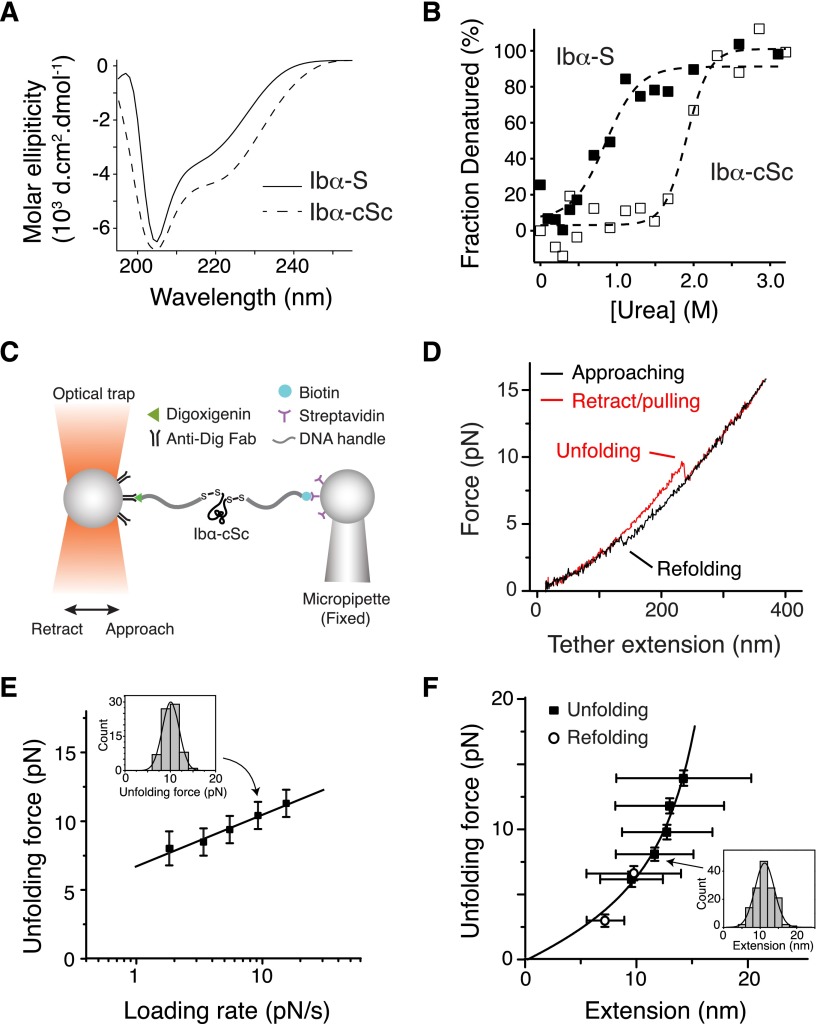
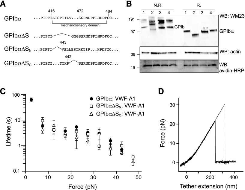
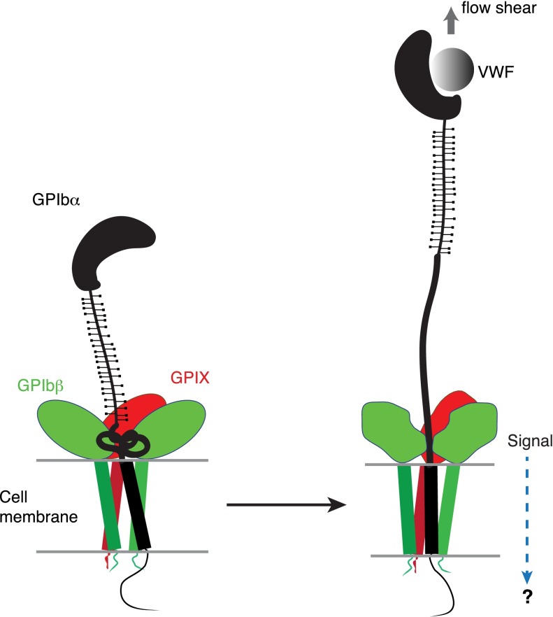
How glycoprotein (GP)Ib-IX complex on the platelet surface senses the blood flow through its binding to the plasma protein von Willebrand factor (VWF) and transmits a signal into the platelet remains unclear. Here we show that optical tweezer-controlled pulling of the A1 domain of VWF (VWF-A1) on GPIb-IX captured by its cytoplasmic domain induced unfolding of a hitherto unidentified structural domain before the dissociation of VWF-A1 from GPIb-IX. Additional studies using recombinant proteins and mutant complexes confirmed its existence in GPIb-IX and enabled localization of this quasi-stable mechanosensitive domain of ∼60 residues between the macroglycopeptide region and the transmembrane helix of the GPIbα subunit. These results suggest that VWF-mediated pulling under fluid shear induces unfolding of the mechanosensitive domain in GPIb-IX, which may possibly contribute to platelet mechanosensing and/or shear resistance of VWF-platelet interaction. The identification of the mechanosensitive domain in GPIb-IX has significant implications for the pathogenesis and treatment of related blood diseases.


Figures

Figure 1
Figure 1
Pulling the engaged A1 domain of VWF induces unfolding of a domain in the full-length GPIb-IX complex. (A) A diagram of GPIb-IX illustrating the experimental setup for the optical tweezer single-molecule force measurement. The BioTag sequence that is specifically recognized and biotinylated by Escherichia coli biotin ligase was either appended to the C-terminus of GPIX cytoplasmic domain (as shown) or placed into the juxtamembrane region of the GPIbα cytoplasmic domain. The biotinylated GPIb-IX was expressed in transfected cells, solubilized in the Triton X-100–containing lysis buffer, and eventually immobilized on the streptavidin-coated bead. Recombinant VWF-A1 was linked through a DNA handle to a polystyrene bead that was placed in an optical trap as described before. Individual domains of GPIbα are marked on the left. (B) The cytoplasmic sequence of GPIX-biotag showing the appended site of biotinylation. A c-myc immunotag and a BioTag sequence (underlined) are attached to the C-terminal end of GPIX. Residues in the GPIX transmembrane domain are marked by a gray box. (C) A single-molecule force-distance trace illustrating the unfolding of MSD before the detachment of VWF-A1 from GPIb-IX. The inset highlights the observed unfolding event.
Figure 2
Figure 2
Quantitation of pulling VWF-A1 from biotinylated GPIb-IX. (A) Plot of lifetimes of the GPIb-IX/VWF-A1 bond vs force (mean ± SEM, n >3). Shown in the insert is a representative histogram of unbinding force (collected under a pulling speed of 100 nm/s) used to obtain bond lifetimes. (B) Fit of unfolding force vs extension data to the WLC model (dashed line), which yielded a contour length of 25.1 ± 0.3 nm. Extension distances were sorted by unfolding force into 4-pN bins. A histogram of extension (insert) was used to find peak extension. Unfolding forces were averaged for each bin (n = 23-51 per bin). Error bars are 1 SD for force and half-bin width for extension.
Figure 3
Figure 3
Localization of MSD in the juxtamembrane stalk region of GPIbα. (A) Overlaid force-distance traces of pulling VWF-A1 on GPIb-IX captured by biotinylated WM23 (red trace) and on GPIb-IX captured by biotin at the GPIX cytoplasmic domain (gray). (B) Plots of lifetimes of the GPIb-IX/VWF-A1 bond vs force (mean ± SEM, n >3).
Figure 4
Figure 4
Unfolding of the recombinant stalk region of GPIbα. (A) Overlaid CD spectra of Ibα-S (solid trace) and Ibα-cSc (dashed) in 50 mM Tris, 50 mM NaCl, and 1 mM DTT; pH 7.4 buffer at 20°C. (B) Chemical denaturation plots of Ibα-S (filled squares) and Ibα-cSc (open squares). (C) Illustration of the optical tweezer setup to measure force-induced unfolding of Ibα-cSc. (D) Force-distance traces showing the force-induced unfolding and refolding of Ibα-cSc from pulling at 100 nm/s. (E) Plot of most probable unfolding force as a function of loading rate. Unfolding forces at 5 different loading rates were plotted as histograms (inset). Each histogram was fitted to a Gaussian curve (inset, solid line) to obtain the most probable force. Uncertainty in force is shown as half of the bin width. The solid line is a linear fit of the data to the Bell-Evans model. (F) Unfolding force-extension data (black squares) fitted to the WLC model, yielding a contour length of 22.3 ± 0.2 nm. Extension distances were sorted by unfolding force into 2-pN bins. A histogram of extension of each bin (inset) was fitted to a Gaussian curve (inset, solid line) to find peak extension. Unfolding forces were averaged for each bin (n = 43-149 per bin for unfolding, and 40-64 per bin for refolding). Error bars are 1 SD for force and half-width of the Gaussian fit for extension. Open circles represent refolding force-shortening data, which were treated the same way as the unfolding forces-extension.
Figure 5
Figure 5
Lack of force-induced unfolding in GPIb-IX complexes with altered MSD. (A) Sequences illustrating various deletion mutations in MSD. (B) Expression and assembly of the mutant complexes, as shown by Western blots under nonreducing (N.R.) or reducing (R.) conditions. Lane 1, cells expressing wild-type complex GPIbα/GPIbβ/GPIX; lane 2, GPIbα∆S/GPIbβ/GPIX; lane 3, GPIbα∆SN/GPIbβ/GPIX; lane 4, GPIbα∆SC/GPIbβ/GPIX. (C) Lifetimes (mean ± SEM of >3 experiments) of the bonds between VWF-A1 and the 3 constructs as a function of force. Each plot is identified by the identity of GPIbα subunit in the complex and the pulling ligand. (D) Representative force-distance traces of pulling VWF-A1 on GPIbα∆SN/GPIbβ/GPIX (thick trace) or GPIbα∆SC/GPIbβ/GPIX (thin trace) complex that was captured by biotin at the GPIX cytoplasmic, showing the lack of force-induced unfolding.
Figure 6
Figure 6
A model of the proposed mechanosensing mechanism of GPIb-IX. We found in this study that the juxtamembrane MSD in GPIbα is folded, and it unfolds upon VWF-mediated pulling. Our results suggest that on the cell surface, the juxtamembrane MSD in GPIbα is folded in the absence of shear flow (left panel). VWF binding under shear to the N-terminal domain of GPIbα induces unfolding of MSD, and subsequently a conformational change in the adjacent extracellular domains of GPIbβ and GPIX, which sends in a signal across the platelet membrane (right).

Comment in

References

    1. Moake JL, Turner NA, Stathopoulos NA, Nolasco LH, Hellums JD. Involvement of large plasma von Willebrand factor (vWF) multimers and unusually large vWF forms derived from endothelial cells in shear stress-induced platelet aggregation. J Clin Invest. 1986;78(6):1456–1461. - PMC - PubMed
    1. Peterson DM, Stathopoulos NA, Giorgio TD, Hellums JD, Moake JL. Shear-induced platelet aggregation requires von Willebrand factor and platelet membrane glycoproteins Ib and IIb-IIIa. Blood. 1987;69(2):625–628. - PubMed
    1. Zhang X, Halvorsen K, Zhang CZ, Wong WP, Springer TA. Mechanoenzymatic cleavage of the ultralarge vascular protein von Willebrand factor. Science. 2009;324(5932):1330–1334. - PMC - PubMed
    1. Auton M, Sowa KE, Behymer M, Cruz MA. N-terminal flanking region of A1 domain in von Willebrand factor stabilizes structure of A1A2A3 complex and modulates platelet activation under shear stress. J Biol Chem. 2012;287(18):14579–14585. - PMC - PubMed
    1. Tischer A, Madde P, Blancas-Mejia LM, Auton M. A molten globule intermediate of the von Willebrand factor A1 domain firmly tethers platelets under shear flow. Proteins. 2014;82(5):867–878. - PMC - PubMed

Publication types

MeSH terms

Substances