

An optimized disaggregation method for human lung tumors that preserves the phenotype and function of the immune cells
- PMID: 25359999
- PMCID: PMC4771617
- DOI: 10.1189/jlb.5TA0814-373
An optimized disaggregation method for human lung tumors that preserves the phenotype and function of the immune cells
Abstract
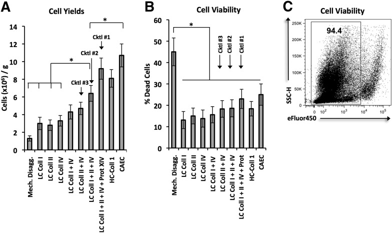
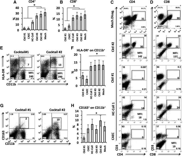
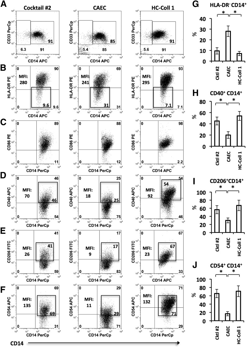
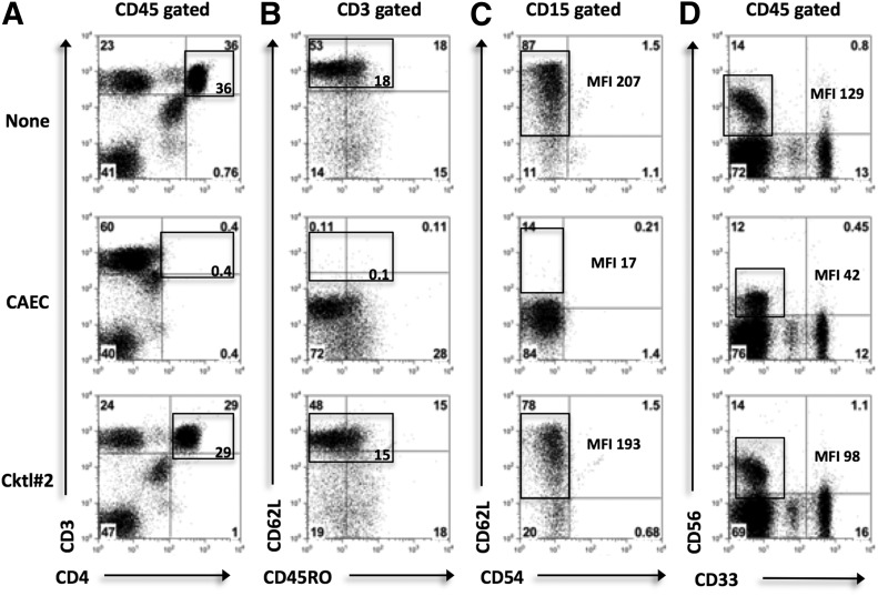
Careful preparation of human tissues is the cornerstone of obtaining accurate data in immunologic studies. Despite the essential importance of tissue processing in tumor immunology and clinical medicine, current methods of tissue disaggregation have not been rigorously tested for data fidelity. Thus, we critically evaluated the current techniques available in the literature that are used to prepare human lung tumors for immunologic studies. We discovered that these approaches are successful at digesting cellular attachments and ECMs; however, these methods frequently alter the immune cell composition and/or expression of surface molecules. We thus developed a novel approach to prepare human lung tumors for immunologic studies by combining gentle mechanical manipulation with an optimized cocktail of enzymes used at low doses. This enzymatic digestion cocktail optimized cell yield and cell viability, retrieved all major tumor-associated cell populations, and maintained the expression of cell-surface markers for lineage definition and in vivo effector functions. To our knowledge, we present the first rigorously tested disaggregation method designed for human lung tumors.
Keywords: collagenase; enzymatic digestion; tumor-infiltrating immune cells.
© Society for Leukocyte Biology.
Figures

Similar articles
-
Phenotypic characterization and functional analysis of human tumor immune infiltration after mechanical and enzymatic disaggregation.J Immunol Methods. 2011 Sep 30;372(1-2):119-26. doi: 10.1016/j.jim.2011.07.002. Epub 2011 Jul 18. J Immunol Methods. 2011. PMID: 21782822
-
Handling, storage, and preparation of human tissues.Curr Protoc Cytom. 2001 May;Chapter 5:Unit 5.2. doi: 10.1002/0471142956.cy0502s01. Curr Protoc Cytom. 2001. PMID: 18770712
-
Immune Cell Composition in Human Non-small Cell Lung Cancer.Front Immunol. 2019 Feb 1;9:3101. doi: 10.3389/fimmu.2018.03101. eCollection 2018. Front Immunol. 2019. PMID: 30774636 Free PMC article.
-
Optimal molecular profiling of tissue and tissue components: defining the best processing and microdissection methods for biomedical applications.Mol Biotechnol. 2005 Feb;29(2):119-52. doi: 10.1385/MB:29:2:119. Mol Biotechnol. 2005. PMID: 15699569 Review.
-
Flow cytometry as an analytical and preparative tool in immunology.J Immunol Methods. 1982;50(3):R85-112. doi: 10.1016/0022-1759(82)90161-2. J Immunol Methods. 1982. PMID: 7047643 Review. No abstract available.
Cited by
-
Capabilities of Single Cell ICP-MS for the Analysis of Cell Suspensions from Solid Tissues.Nanomaterials (Basel). 2022 Dec 20;13(1):12. doi: 10.3390/nano13010012. Nanomaterials (Basel). 2022. PMID: 36615921 Free PMC article.
-
Phenotypic and functional analysis of malignant mesothelioma tumor-infiltrating lymphocytes.Oncoimmunology. 2019 Jul 13;8(9):e1638211. doi: 10.1080/2162402X.2019.1638211. eCollection 2019. Oncoimmunology. 2019. PMID: 31428531 Free PMC article.
-
Folate Receptor Beta Designates Immunosuppressive Tumor-Associated Myeloid Cells That Can Be Reprogrammed with Folate-Targeted Drugs.Cancer Res. 2021 Feb 1;81(3):671-684. doi: 10.1158/0008-5472.CAN-20-1414. Epub 2020 Nov 17. Cancer Res. 2021. PMID: 33203700 Free PMC article.
-
Intra-tumoral delivery of CXCL11 via a vaccinia virus, but not by modified T cells, enhances the efficacy of adoptive T cell therapy and vaccines.Oncoimmunology. 2018 Jan 9;7(3):e1395997. doi: 10.1080/2162402X.2017.1395997. eCollection 2018. Oncoimmunology. 2018. PMID: 29399394 Free PMC article.
-
Phenotype and function of tumor-associated neutrophils and their subsets in early-stage human lung cancer.Cancer Immunol Immunother. 2017 Aug;66(8):997-1006. doi: 10.1007/s00262-017-1976-0. Epub 2017 Mar 10. Cancer Immunol Immunother. 2017. PMID: 28283697 Free PMC article. Review.
References
-
- Visscher D. W., Crissman J. D. (1994) Dissociation of intact cells from tumors and normal tissues. In Methods in Cell Biology (Darzynkiewicz Z., Robinson J. P., Crissman H. A., eds.), Academic Press, San Diego, CA, USA, 1–13. - PubMed
-
- Slocum H. K., Pavelic Z. P., Rustum Y. M., Creaven P. J., Karakousis C., Takita H., Greco W. R. (1981) Characterization of cells obtained by mechanical and enzymatic means from human melanoma, sarcoma, and lung tumors. Cancer Res. 41, 1428–1434. - PubMed
-
- Grange C., Létourneau J., Forget M. A., Godin-Ethier J., Martin J., Liberman M., Latour M., Widmer H., Lattouf J. B., Piccirillo C. A., Cailhier J. F., Lapointe R. (2011) Phenotypic characterization and functional analysis of human tumor immune infiltration after mechanical and enzymatic disaggregation. J. Immunol. Methods 372, 119–126. - PubMed
-
- Petit V., Massonnet G., Maciorowski Z., Touhami J., Thuleau A., Némati F., Laval J., Château-Joubert S., Servely J. L., Vallerand D., Fontaine J. J., Taylor N., Battini J. L., Sitbon M., Decaudin D. (2013) Optimization of tumor xenograft dissociation for the profiling of cell surface markers and nutrient transporters. Lab. Invest. 93, 611–621. - PubMed
Publication types
MeSH terms
Grants and funding
LinkOut - more resources
Full Text Sources
Other Literature Sources
Medical
