. 2014 Jan:46:591-614.
doi: 10.1146/annurev-fluid-010313-141309.

Fluid Mechanics, Arterial Disease, and Gene Expression

Affiliations

Fluid Mechanics, Arterial Disease, and Gene Expression

John M Tarbell et al. Annu Rev Fluid Mech. 2014 Jan.

Abstract

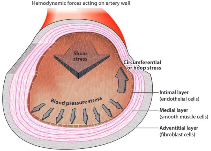
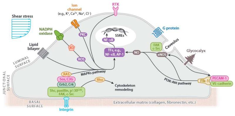
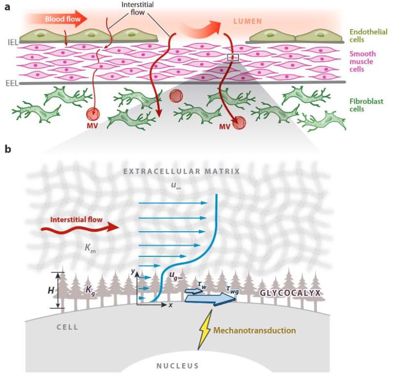
This review places modern research developments in vascular mechanobiology in the context of hemodynamic phenomena in the cardiovascular system and the discrete localization of vascular disease. The modern origins of this field are traced, beginning in the 1960s when associations between flow characteristics, particularly blood flow-induced wall shear stress, and the localization of atherosclerotic plaques were uncovered, and continuing to fluid shear stress effects on the vascular lining endothelial) cells (ECs), including their effects on EC morphology, biochemical production, and gene expression. The earliest single-gene studies and genome-wide analyses are considered. The final section moves from the ECs lining the vessel wall to the smooth muscle cells and fibroblasts within the wall that are fluid me chanically activated by interstitial flow that imposes shear stresses on their surfaces comparable with those of flowing blood on EC surfaces. Interstitial flow stimulates biochemical production and gene expression, much like blood flow on ECs.

Keywords: endothelial cells; glycocalyx; interstitial flow; mechanotransduction; shear stress; smooth muscle cells.


Figures

Figure 1
Figure 1
The arterial wall consists of three layers: the intima, the innermost layer that is in contact with blood, which consists of a lining layer of endothelial cells and a thin basement matrix for endothelial attachment; the media, which contains layers of smooth muscle cells that are separated by elastic lamina; and the adventitia, the outermost layer, which contains fibroblasts embedded in loose connective tissue. The forces (stresses) acting on the arterial wall are the normal stress of blood pressure that is balanced by the circumferential stress in the wall and the fluid shear stress that is tangential to the endothelial cell surface.
Figure 2
Figure 2
The eight stages of development of an atherosclerotic plaque. First LDL moves into the subendothelium and is oxidized by Macrophages and smooth muscle cells (steps 1 and 2). The release of growth factors and cytokines attracts additional monocytes (steps 3 and 4). Foam cell accumulation and smooth muscle cell proliferation result in the growth of the plaque (steps 6–8). Figure adapted with permission from Faxon et al. (2004).
Figure 3
Figure 3
Hemodynamics in the human carotid bifurcation display a wide range of wall shear stress (WSS) characteristics. (Left.) The carotid anatomy of a normal human subject (27-year-old male) was reconstructed from noninvasive magnetic resonance imaging measurements. The color-coded map shown in this carotid model displays the time-averaged WSS magnitude at different points along the vascular wall. Clearly the carotid sinus is a region of very low mean WSS and also a site of atherosclerotic plaque development. Regions in the distal internal carotid artery and carotid sinus were selected, and the time-dependent WSS values along the mean flow direction from multiple individual points within each region were plotted for one cardiat cycle (right). For the carotid sinus, the peak WSS clearly is lower than in the distal carotid, and the minimum WSS in the carotid is actually negative, indicating a time interval of WSS reversal (retrograde flow) that is not apparent in the distal carotid. Figure adapted with permission from Dai et al. (2004).
Figure 4
Figure 4
Mechanotransduction signaling pathways in endothelial cells in response to (a) laminar or (b) oscillatory shear stress stimuli. Laminar shear stress stabilizes a healthy, anti-inflammatory state in endothelial cells that results in cytoskeletal remodeling and endothelial cell alignment in the direction of flow, as well as increased nitric oxide production and suppresion of inflammatory cell adhesion molecules. Conversely, disturbed flow or oscillatory shear stress induces an inflammatory and thrombotic state characterized by high expression of cell adhesion molecules and production of inflammatory cytokines, high oxidative stress, lack of cell alignment with flow, and a leaky endothelial cell barrier that allows infiltration of smooth muscle cells and immunomodulatory cell types (Noguchi & Jo 2011).
Figure 5
Figure 5
Mechanosensory pathways utilizing surface mechanoreceptors [e.g., ion channels (K+, Ca2+, Na+, Cl), receptor tyrosine kinases, G protein coupled receptors), cell-cell and cell-matrix adhesion complexes (e.g., PECAM-1/VE-cadherin/VEGFR2 complex and focal adhesion kinases), the glycocalyx, and cytoskeletal elements that transduce signals and result in downstream signal amplification via master regulators such as kinase cascades and transcription factor networks. These signaling events alter the endothelial cell phenotype and behavior in response to extracellular biomechanical stimuli. Figure adapted with permission from Chatzizisis et al. (2007).
Figure 6
Figure 6
Interstitial flow shear stress and vascular injury. (a) Vascular smooth muscle cells (SMCs) and fibroblasts (FBs) in the intact artery are not exposed to luminal blood flow shear stress that normally is experienced by endothelial cells but are exposed to a very low physiological transmural interstitial flow (top left). The interstitial flow is driven by the pressure diffrentials between the arterial lumen and the adventital microvessels (MVs) (i.e., vasa vasorum and lymphatics) or the surrounding tissue. After endothelial denudation, the superficial SMCs may be exposed to the blood flow, and the medial SMCs and adventitial FBs are exposed to elevated interstitial fluid flow. (b) The glycocalyx can mediate interstitial flow mechanotransduction by sensing interstitial flow and transmitting solid stresses (τwg) to the cell membrane that are 10–100-fold higher than the fluid shear stress (τw)τwgτwH(Kg) H is the glycocalyx thickness Kg the Darcy permeability of the glycocalyx, Km the Darcy permeability of the surrounding extracellular matrix, μ the superficial velocity far from the surface, and μg the velocity in the glycocalyx layer far from the cell surface. Readers are referred to Wang & Tarbell (1995) and Tarbell & Shi (2013) for detailed modeling. Figure adapted with permission from Shi & Tarbell (2011) and Tarbell & Shi (2013). Abbreviations: EEL, external elastc lamina; IEL, internal elastic lamina.

Similar articles

Cited by

References

    1. Abumiya T, Sasaguri T, Taba Y, Miwa Y, Miyagi M. Shear stress induces expression of vascular endothelial growth factor receptor Flk-1/KDR through the CT-rich Sp1 binding site. Arterioscler. Thromb. Vasc. Biol. 2002;22:907–13. - PubMed
    1. Ainslie KM, Garanich JS, Dull RO, Tarbell JM. Vascular smooth muscle cell glycocalyx influences shear stress-mediated contractile response. J. Appl. Physiol. 2005;98:242–49. - PubMed
    1. Akimoto S, Mitsumata M, Sasaguri T, Yoshida Y. Laminar shear stress inhibits vascular endothelial cell proliferation by inducing cyclin-dependent kinase inhibitor p21Sdi1/Cip1/Waf1. Circ. Res. 2000;86:185–90. - PubMed
    1. Alshihabi SN, Chang YS, Frangos JA, Tarbell JM. Shear stress-induced release of PGE2 and PGI2 by vascular smooth muscle cells. Biochem. Biophys. Res. Commun. 1996;224:808–14. - PubMed
    1. Apenberg S, Freyberg MA, Friedl P. Shear stress induces apoptosis in vascular smooth muscle cells via an autocrine Fas/FasL pathway. Biochem. Biophys. Res. Commun. 2003;310:355–59. - PubMed