. 2014 Oct 15:8:198.
doi: 10.3389/fnsys.2014.00198. eCollection 2014.

Differential loss of thalamostriatal and corticostriatal input to striatal projection neuron types prior to overt motor symptoms in the Q140 knock-in mouse model of Huntington's disease

Affiliations

Differential loss of thalamostriatal and corticostriatal input to striatal projection neuron types prior to overt motor symptoms in the Q140 knock-in mouse model of Huntington's disease

Yun-Ping Deng et al. Front Syst Neurosci. .

Abstract

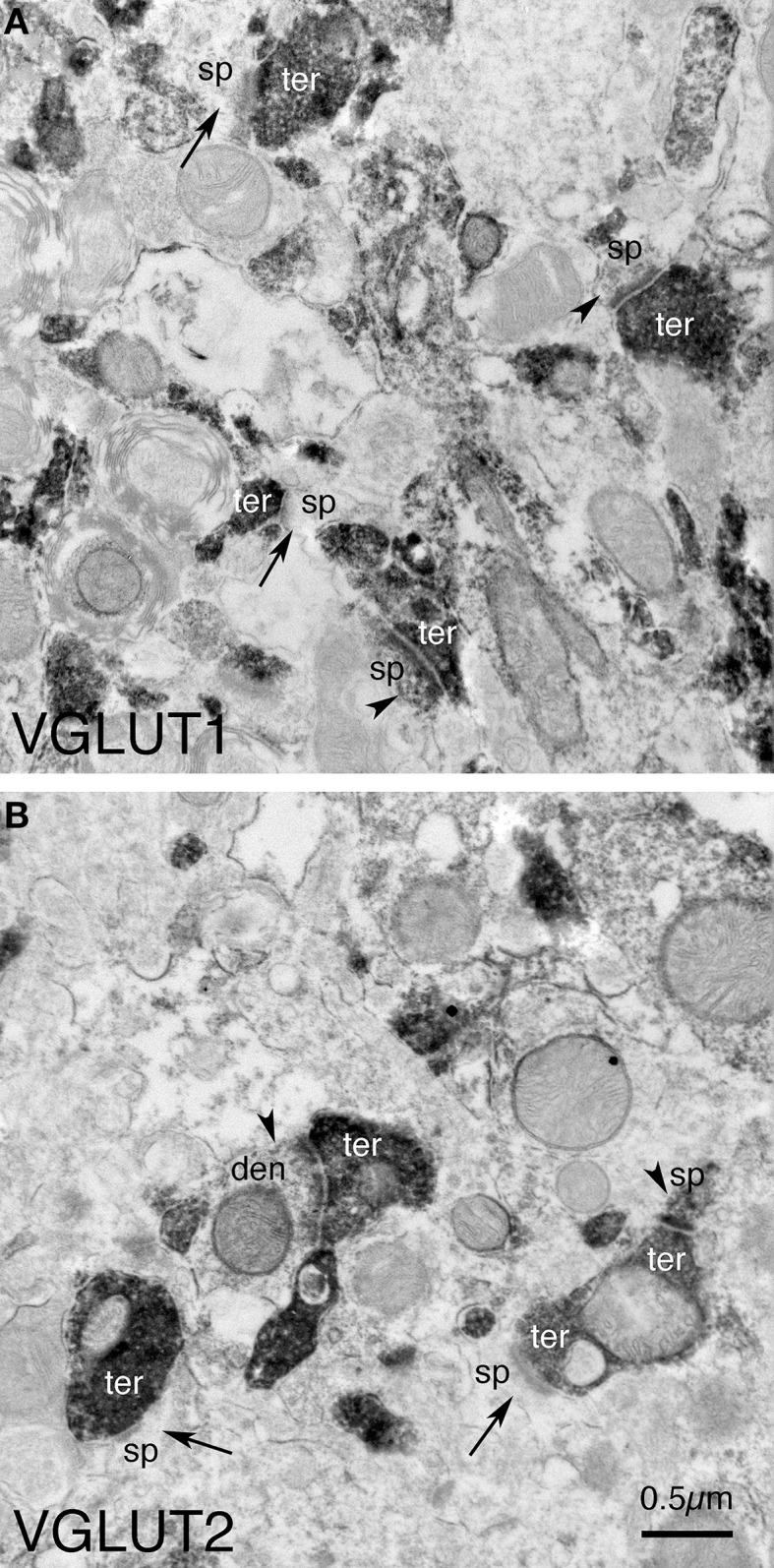
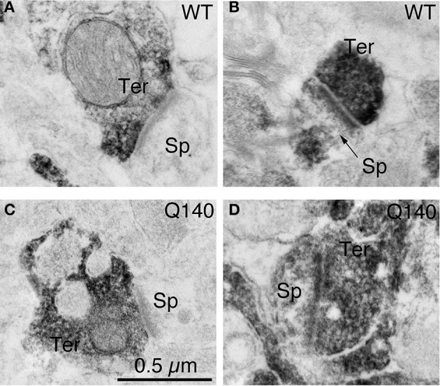
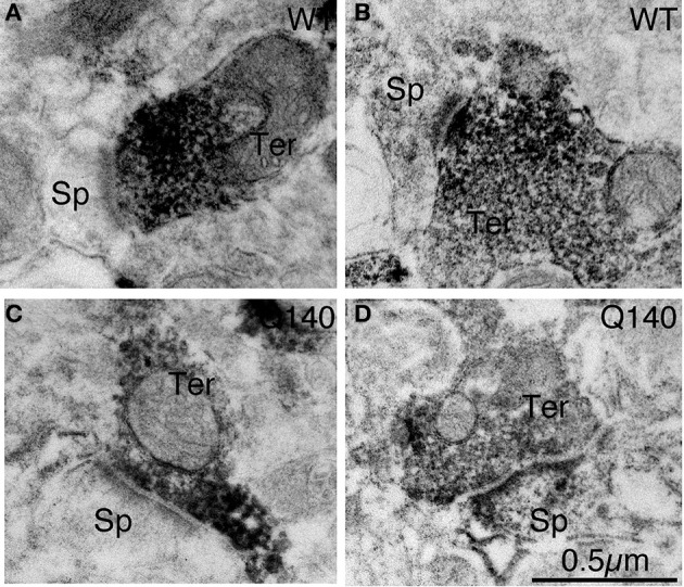
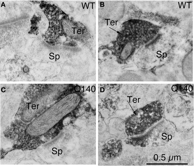
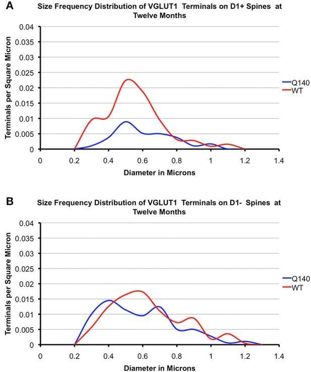
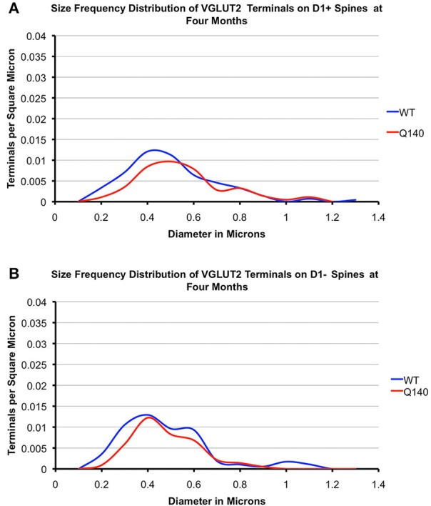
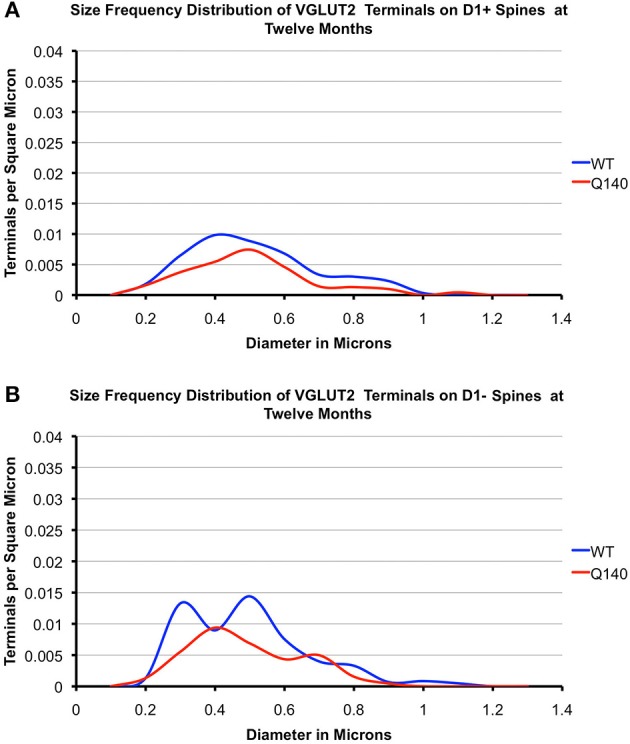
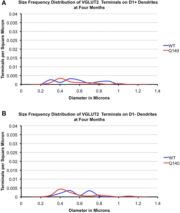
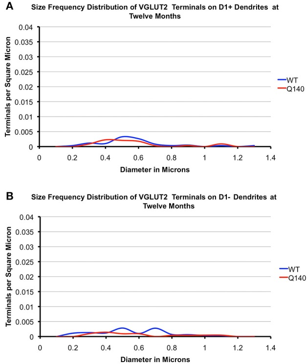
Motor slowing and forebrain white matter loss have been reported in premanifest Huntington's disease (HD) prior to substantial striatal neuron loss. These findings raise the possibility that early motor defects in HD may be related to loss of excitatory input to striatum. In a prior study, we showed that in the heterozygous Q140 knock-in mouse model of HD that loss of thalamostriatal axospinous terminals is evident by 4 months, and loss of corticostriatal axospinous terminals is evident at 12 months, before striatal projection neuron pathology. In the present study, we specifically characterized the loss of thalamostriatal and corticostriatal terminals on direct (dSPN) and indirect (iSPN) pathway striatal projection neurons, using immunolabeling to identify thalamostriatal (VGLUT2+) and corticostriatal (VGLUT1+) axospinous terminals, and D1 receptor immunolabeling to distinguish dSPN (D1+) and iSPN (D1-) synaptic targets. We found that the loss of corticostriatal terminals at 12 months of age was preferential for D1+ spines, and especially involved smaller terminals, presumptively of the intratelencephalically projecting (IT) type. By contrast, indirect pathway D1- spines showed little loss of axospinous terminals at the same age. Thalamostriatal terminal loss was comparable for D1+ and D1- spines at both 4 and 12 months. Regression analysis showed that the loss of VGLUT1+ terminals on D1+ spines was correlated with a slight decline in open field motor parameters at 12 months. Our overall results raise the possibility that differential thalamic and cortical input loss to SPNs is an early event in human HD, with cortical loss to dSPNs in particular contributing to premanifest motor slowing.

Keywords: Huntington's disease; corticostriatal; pathology; premanifest; thalamostriatal.


Figures

Figure 1
Figure 1
Examples of the fields of view captured in the EM images used for analysis. Image (A) shows VGLUT1+ immunolabeled synaptic terminals (ter) on D1+ (arrowheads) and D1− (arrows) spines (sp) and dendrites (den) in striatum in WT mice at 12 months of age. Image (B) shows VGLUT2+ immunolabeled synaptic terminals (ter) on D1+ (arrowheads) and D1− (arrows) spines (sp) and dendrites (den) in striatum in WT mice at 12 months of age. Both images are at the same magnification.
Figure 2
Figure 2
Examples of EM images of VGLUT1+ immunolabeled synaptic terminals on D1− (A) and D1+ (B) spines in striatum in WT mice at 12 months of age, and of VGLUT1+ immunolabeled synaptic terminals on D1− (C) and D1+ (D) spines in striatum in Q140 mice at 12 months of age. All images are at the same magnification.
Figure 3
Figure 3
Examples of EM images of VGLUT2+ immunolabeled synaptic terminals on D1− (A) and D1+ (B) spines in striatum in WT mice at 4 months of age, and of VGLUT2+ immunolabeled synaptic terminals on D1− (C) and D1+ (D) spines in striatum in Q140 mice at 4 months of age. All images are at the same magnification.
Figure 4
Figure 4
Examples of EM images of VGLUT2+ immunolabeled synaptic terminals on D1− (A) and D1+ (B) spines in striatum in WT mice at 12 months of age, and of VGLUT2+ immunolabeled synaptic terminals on D1− (C) and D1+ (D) spines in striatum in Q140 mice at 12 months of age. All images are at the same magnification.
Figure 5
Figure 5
Graphs showing the size frequency distributions for VGLUT1+ axospinous synaptic terminals on D1+ (A) and D1− (B) striatal projection neurons in striatum of 12 month-old WT and heterozygous Q140 mice. Note that the large shortfall in small terminals on D1+ spines in Q140 mice.
Figure 6
Figure 6
Graphs showing the size frequency distributions for VGLUT2+ axospinous synaptic terminals on D1+ (A) and D1− (B) striatal projection neurons in striatum of 4 month-old WT and heterozygous Q140 mice. Note that the shortfall in VGUT2+ axospinous terminals on both D1+ and D1− spines in Q140 mice.
Figure 7
Figure 7
Graphs showing the size frequency distributions for VGLUT2+ axospinous synaptic terminals on D1+ (A) and D1− (B) striatal projection neurons in striatum of 12 month-old WT and heterozygous Q140 mice. Note that the shortfall in VGUT2+ axospinous terminals on both D1+ and D1− spines in Q140 mice.
Figure 8
Figure 8
Graphs showing the size frequency distributions for VGLUT2+ axodendritic synaptic terminals on D1+ (A) and D1− (B) striatal projection neurons in striatum of 4 month-old WT and heterozygous Q140 mice. Note that VGUT2+ axodendritic terminals on both D1+ and D1− spines are largely similar in abundance in WT and Q140 mice, but with some possible shortfall in large terminals on D1− dendrites.
Figure 9
Figure 9
Graphs showing the size frequency distributions for VGLUT2+ axodendritic synaptic terminals on D1+ (A) and D1− (B) striatal projection neurons in striatum of 12 month-old WT and heterozygous Q140 mice. Note that VGUT2+ axodendritic terminals on both D1+ and D1− spines are largely similar in abundance in WT and Q140 mice, but with some possible shortfall in large terminals on D1− dendrites.

Similar articles

Cited by

References

    1. Aggerman K., Ernfors P. (2003). Differential influence of BDNF and NT3 on the expression of calcium binding proteins and neuropeptide Y in vivo. Neuroreport 14, 2183–2187 10.1097/00001756-200312020-00010 - DOI - PubMed
    1. Albin R. L., Qin Y., Young A. B., Penney J. B., Chesselet M. F. (1991). Preproenkephalin messenger RNA-containing neurons in striatum of patients with symptomatic and presymptomatic Huntington's disease: an in situ hybridization study. Ann. Neurol. 30, 542–549 10.1002/ana.410300406 - DOI - PubMed
    1. Albin R. L., Young A. B., Penney J. B. (1989). The functional anatomy of basal ganglia disorders. Trends Neurosci. 12, 366–375 10.1016/0166-2236(89)90074-X - DOI - PubMed
    1. Alcantara S., Frisen J., del Rio J. A., Soriano E., Barbacid M., Silos-Santiago I. (1997). TrkB signaling is required for postnatal survival of CNS neurons and protects hippocampal and motor neurons from axotomy-induced cell death. J. Neurosci. 17, 3623–3633 - PMC - PubMed
    1. André V. M., Cepeda C., Fisher Y. E., Huynh M., Bardakjian N., Singh S., et al. (2011a). Differential electrophysiological changes in striatal output neurons in Huntington's disease. J. Neurosci. 31, 1170–1182 10.1523/JNEUROSCI.3539-10.2011 - DOI - PMC - PubMed

LinkOut - more resources