Review
. 2014 Oct 16:5:546.
doi: 10.3389/fpls.2014.00546. eCollection 2014.

Analogous reserve distribution and tissue characteristics in quinoa and grass seeds suggest convergent evolution

Affiliations
Review

Analogous reserve distribution and tissue characteristics in quinoa and grass seeds suggest convergent evolution

Hernán P Burrieza et al. Front Plant Sci. .

Abstract

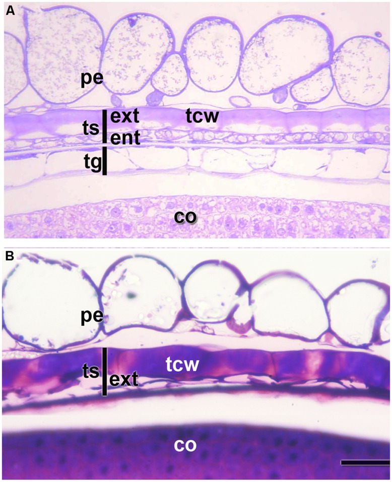
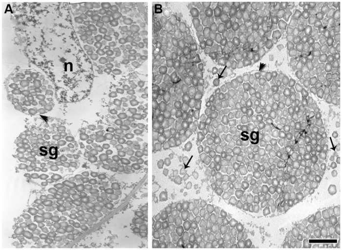
Quinoa seeds are highly nutritious due to the quality of their proteins and lipids and the wide range of minerals and vitamins they store. Three compartments can be distinguished within the mature seed: embryo, endosperm, and perisperm. The distribution of main storage reserves is clearly different in those areas: the embryo and endosperm store proteins, lipids, and minerals, and the perisperm stores starch. Tissues equivalent (but not homologous) to those found in grasses can be identified in quinoa, suggesting the effectiveness of this seed reserve distribution strategy; as in cells of grass starchy endosperm, the cells of the quinoa perisperm endoreduplicate, increase in size, synthesize starch, and die during development. In addition, both systems present an extra-embryonic tissue that stores proteins, lipids and minerals: in gramineae, the aleurone layer(s) of the endosperm; in quinoa, the micropylar endosperm; in both cases, the tissues are living. Moreover, the quinoa micropylar endosperm and the coleorhiza in grasses play similar roles, protecting the root in the quiescent seed and controlling dormancy during germination. This investigation is just the beginning of a broader and comparative study of the development of quinoa and grass seeds. Several questions arise from this study, such as: how are synthesis and activation of seed proteins and enzymes regulated during development and germination, what are the genes involved in these processes, and lastly, what is the genetic foundation justifying the analogy to grasses.

Keywords: coleorhiza; endosperm; grass seed; micropylar endosperm; perisperm; quinoa seed.


Figures

FIGURE 1
FIGURE 1
(A) Quinoa grain; (B) Quinoa seed (without pericarp); (C) Longitudinal midsection of a quinoa seed; (D) Excised embryo. ax, hypocotyl-radicle axis; co, cotyledon; ps, perisperm; ram, root apical meristem. Bar: 1 mm.
FIGURE 2
FIGURE 2
Pericarp and integuments at two stages of quinoa seed development. (A) Young seed (torpedo stage). (B) Mature seed. Grains were fixed using a mixture of 2% paraformaldehyde and 1% glutaraldehyde in 0.1 M phosphate buffer, pH 7.2, and embedded in London Resin White resin, according to López-Fernández and Maldonado (b). Semithin sections (2 μm thick) were stained with 0.5 % Toluidine Blue. co, cotyledon; ent, endotesta; ext, exotesta; pe, pericarp; tcw, tangential cell wall of the cells from the outer layer of the outer integument; tg, tegmen; ts, testa.
FIGURE 3
FIGURE 3
Quinoa micropylar endosperm (in a mature seed). (A) The white arrow indicates the center of the micropylar cone; co, cotyledon; em, embryo; me, micropylar endosperm; pe, pericarp; ps, perisperm. (B) Detail of (A). The white arrow indicates the central channel of the micropylar cone, which is occupied by the remains of the suspensor. ca, caliptra; me, micropylar endosperm; ram, root apical meristem. Semithin section (2 μm thick) was obtained from a fixed, resin-embedded, sectioned, and stained seed, as described in Figure 2.
FIGURE 4
FIGURE 4
Quinoa micropylar endosperm (at the torpedo stage). cn, crushed nucellus; ee, ephemeral endosperm; le, lasting endosperm; me, micropylar endosperm; su, supensor. Semithin section (2 μm thick) was obtained from a fixed, resin-embedded, sectioned, and stained seed as described in Figure 2. The border between the suspensor and endosperm has been highlighted with a dotted line. White arrows indicate the “festooned” outer cell walls in the transfer cells of the suspensor. Bar: 10 μm.
FIGURE 5
FIGURE 5
Grain development in quinoa (A) and maize (B), from anthesis to maturity. In quinoa, the first image (ovule at anthesis) has been enlarged in the top image. ch, chalazal pole; m, micropylar pole. Bar: 0.5 mm.
FIGURE 6
FIGURE 6
The suspensor in two subsequent early stages of embryo development. (A) Quinoa seed. (B) Grass seed. cl, coleoptile; co, cotyledon primordium; cr, coleorhiza; ram, root apical meristem; sam, shoot apical meristem; s, suspensor.
FIGURE 7
FIGURE 7
Ultrathin sections of the quinoa perisperm in two subsequent developmental stages. Grains were fixed and embedded as described in Figure 2. Ultra-thin sections were mounted on grids coated with Formvar and stained with uranyl acetate followed by lead citrate.(A) Numerous simple starch grains are being packed inside amyloplasts (arrowhead) originating compound grains; n, nucleus; sg, starch grains.(B) Simple starch grains (arrows) and compound starch grains (arrowhead). Bar: 1 μm.
FIGURE 8
FIGURE 8
Origin of protein storage vacuoles (PSVs) and lipid bodies in quinoa embryo. Excised embryos (torpedo stage) were processed as indicated in Figure 7. (A) Cells from the ground meristem. (B) Cells from the procambium. (C) detail of (B). (D–I) Details of cytoplasm and organelles in cells from the ground meristem. In the different images, intense biosynthetic activity in cytoplasm can be inferred by the presence of abundant cisternae of rough endoplasmic reticulum (rer), numerous PSVs originating from the vacuoles (va); globoids (white arrows) can be seen inside PSV; the empy areas inside PSV contained globoid crystals before they were dissolved during treatment of tissue fixation; numerous lipid bodies (lb) associated with endoplasmic reticulum; frequent dictiosomes, or Golgi (gl) and circular vesicles with electronically dense content in proximity to the dictiosomes (see D); nuclei with one or more nucleoli (nu). Arrowheads indicate the presence in the vacuole of the precursor salts of the globoids; white arrows indicate globoids; black arrows indicate plasmodesmata. p, plastid. Bars: A–B, 2 μm; C, 1.5 μm; D–I, 0.5 μm.

References

    1. Amritphale D., Yoneyama K., Takeuchi Y., Ramakrishna P., Kusumoto D. (2005). The modulating effect of the perisperm-endosperm envelope on ABA-inhibition of seed germination in cucumber. J. Exp. Bot. 56 2173–2181. 10.1093/jxb/eri217 - DOI - PubMed
    1. Arana M. V., Burgin M. J., de Miguel L. C., Sánchez R. A. (2007). The very-low-fluence and high-irradiance responses of the phytochromes have antagonistic effects on germination, mannan-degrading activities, and DfGA3ox transcript levels in Datura ferox seeds. J. Exp. Bot. 58 3997–4004. 10.1093/jxb/erm256 - DOI - PubMed
    1. Balzotti M. R. B., Thornton J. N., Maughan P. J., McClellan D. A., Stevens M. R., Jellen E. N., et al. (2008). Expression and evolutionary relationship of the Chenopodium quinoa 11S seed storage protein gene. Int. J. Plant Sci. 169 281–291. 10.1086/523874 - DOI
    1. Barrero J. M., Talbot M. J., White R. G., Jacobsen J. V., Gubler F. (2009). Anatomical and transcriptomic studies of the coleorhiza reveal the importance of this tissue in regulating dormancy in barley. Plant Physiol. 150 1006–1021. 10.1104/pp.109.137901 - DOI - PMC - PubMed
    1. Beckles D. M., Smith A. M., Rees T. (2001). A cytosolic ADP-glucose pyrophosphorylase is a feature of graminaceous endosperms,but not of other starch-storing organs. Plant Physiol. 125 818–827. 10.1104/pp.125.2.818 - DOI - PMC - PubMed

LinkOut - more resources